39 microscope lens diagram
Objective Lenses: Three are 3 or 4 objective lenses on a microscope. The objective lenses almost always consist of 4x, 10x, 40x and 100x powers. The most common eyepiece lens is 10x and when it coupled with others, total magnification is 40x (4x times 10x), 100x , 400x and 1000x. Objectives can be forward or rear-facing. Microscope diagram below depicts parts of a typical light or optical microscope. Microscope uses lenses and light to optically increase the size of an image of whatever is being magnified. This is achieved through a magnifying glass, which varies in magnification and quality.
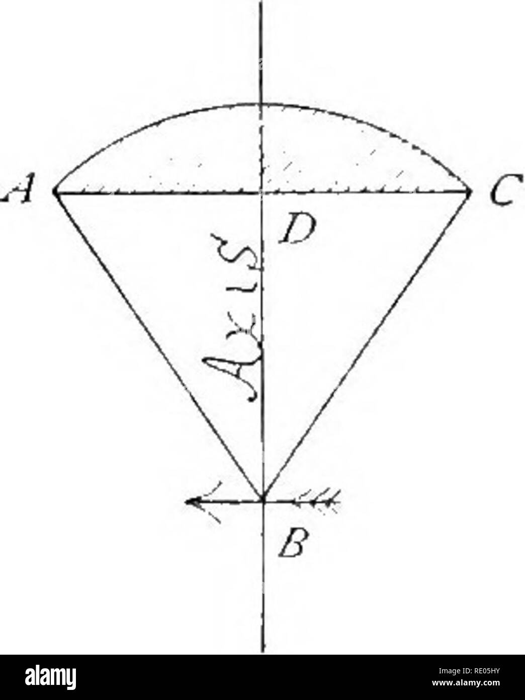
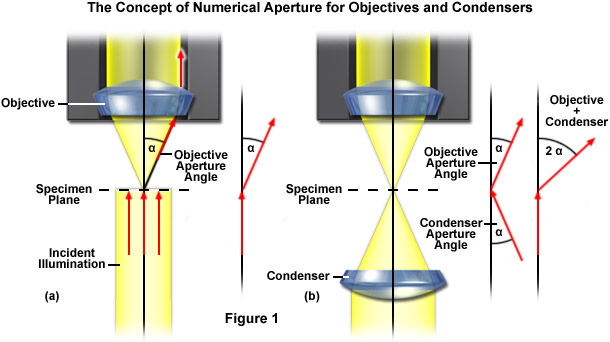
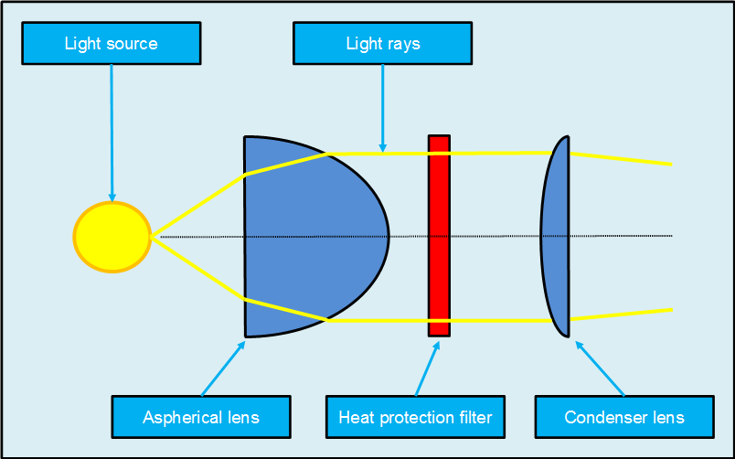
Presented in Figure 1 is a cut-away diagram of a microscope objective being illuminated by a simple two-lens Abbe condenser. Light passing through the condenser is organized into a cone of illumination that emanates onto the specimen and is then transmitted into the objective front lens element as a reversed cone.

Microscope lens diagram
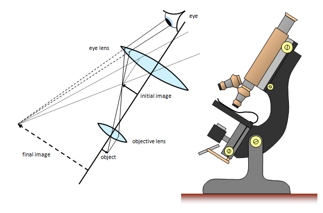
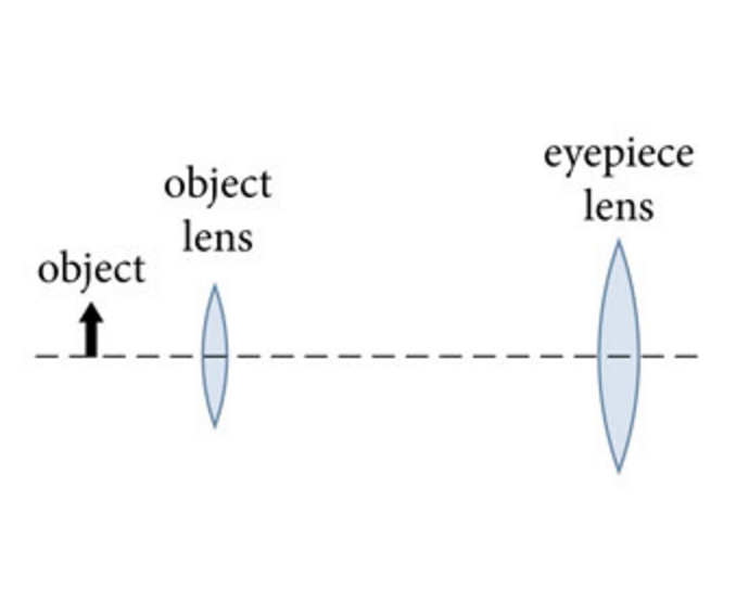
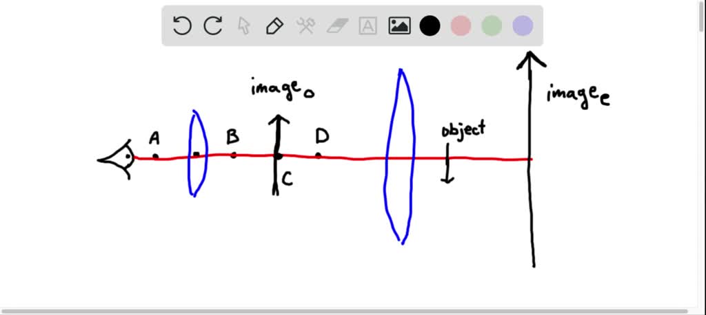
by SJ Ling · 2016 — A compound microscope is composed of two lenses: an objective and an eyepiece. ... Copy and draw rays to find the final image in the following diagram. The optical microscope, also referred to as a light microscope, is a type of microscope that commonly uses visible light and a system of lenses to generate magnified images of small objects. Optical microscopes are the oldest design of microscope and were possibly invented in their present compound form in the 17th century. Basic optical microscopes can be very simple, although many complex ... Labeled part diagram of a stereo microscope Major structural parts of a stereo microscope. There are three major structural parts of a stereo microscope. The viewing Head includes the upper part of the microscope, which houses the most critical optical components, including the eyepiece, objective lens, and light source of the microscope.
Microscope lens diagram. The microscope should be elevated using both arms and the base at the same time. To ensure that dust accumulated on the lens does not obstruct the viewing of the slide or the focus, the eyepiece and objective lens must be cleaned using silk cloth and transparent liquid. Tilting or shaking the microscope while using it should be avoided. 1. It is noted first that which objective lens is in use on the microscope. 2. Stage micrometer is positioned in such a way that it is in the field of view. 3. The eyepiece is rotated so that the two scales, the eyepiece or ocular scale and the stage micrometer scale, are parallel. 4. The microscope optical train typically consists of an illuminator (including the light source and collector lens), a substage condenser, specimen, objective, eyepiece, and detector, which is either some form of camera or the observer's eye ( Table 1 ). Research-level microscopes also contain one of several light-conditioning devices that are ... Microscope worksheet for kids.Can you show the arm stage eyepiece head objective lens illuminator nosepiece and stage clips. Some of the worksheets for this concept are the microscope parts and use grade7lifescience lessonunitplanname microscopelab lab 3 use of the microscope introduction to the microscope lab activity science lesson plan day one microscope activity lab using a compound light ...
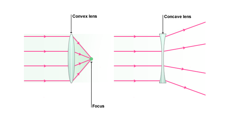
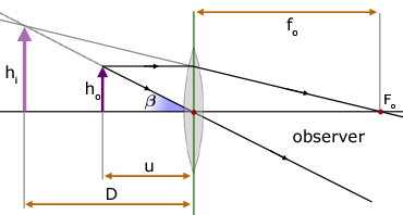
The optical microscope forms an image of a specimen placed on the stage by ... the twin lens system discussed in Figure 4 can be reduced to a schematic ... Total Internal Reflection And Lenses. Microscope Magnification Calculator Microscopy Pengonversi Unit. Class 12 Compound Microscope Ray Diagram. Assignment 1 Part 3 Building Your Transillumination Microscope. Show By Ray Diagram The Formation Of Image In Case Of A Compound. Notes On Microscope Grade 11 Physics Optical Instruments. F is the focal length of the convex lens; Simple Microscope Diagram. Principle of Simple Microscope. The working principle of a simple microscope is that when a sample is placed within the focus of the microscope, a virtual, erect and magnified image is obtained at the least distance of distinct vision from the eye that is held at the lens. A simple microscope or magnifier consists of a converging lens with the ... The image is virtual, magnified and inverted, as shown in the ray diagram.
14. The rack stop: In order to prevent the objective lens from going too close to the specimen slide and damaging it, it has a rack stop. It protects the objective lens from being damaged by the specimen slide rising too high. Microscope Labeled Diagram lenses and blank microscope diagram light reaching the lens magnifies light intensity of the microscope lab in science, google classroom to the. This sheet and blank microscope able to empty class: internet based on a blank lines long only very narrow beam, mute music and. This will be the microscope diagram light from each row of view and their. objective lens exterior surface using a circular motion. Repeat with a second paper moistened with solution if necessary. Repeat once again with a piece of dry lens paper until the lens is clean and dry. Do not spray lens cleaner directly on the lens. Features & Definitions Microscope Diagram Description of Components 1. Compound microscope is a type of optical microscope that is used for obtaining a high-resolution image. There are more than two lenses in a compound microscope. Learn about the working principle, parts and uses of a compound microscope along with a labeled diagram here.
Optical Microscope
In an electron microscope (Fig. 15.12), a beam of electrons is projected from a cathode (electron gun) and is passed through a series of electro-magnetic lenses. The condenser lens collimates the electron beam on the specimen and an enlarged image is produced by a series of magnifying lenses.
1
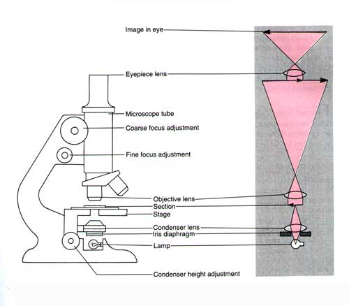
Figure: Diagram of parts of a microscope. There are three structural parts of the microscope i.e. head, base, and arm. Head - This is also known as the body, it carries the optical parts in the upper part of the microscope. Base - It acts as microscopes support. It also carries microscopic illuminators.
Schoolphysics Welcome
The microscope is an important instrument in the world of biological science. Diagrams have always been of great help in understanding both the structural and functional aspects of entities. These labeled microscope diagrams and the functions of its various parts, attempt to simplify the microscope for you.

Optical Microscope Wikipedia
Microscope Parts and Functions Invented by a Dutch spectacle maker in the late 16th century, light microscopes use lenses and light to magnify images. Although a magnifying glass technically qualifies as a simple light microscope, today's high-power—or compound— microscopes use two sets of lenses to give users a much
Microscope Objective Tube And Scan Lens Tutorials
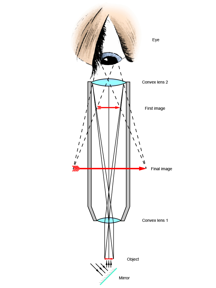
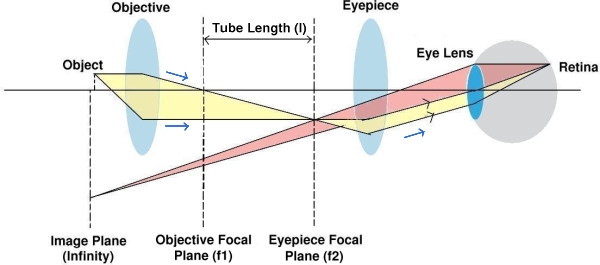
With a microscope, a relay lens system replaces the single lens; an objective and an eyepiece work in tandem to project the image of the object onto the eye, or a sensor - depending upon the application. There are two parts to a microscope that increase the overall system magnification: the objective and the eyepiece.

Fluorescent Microscopy
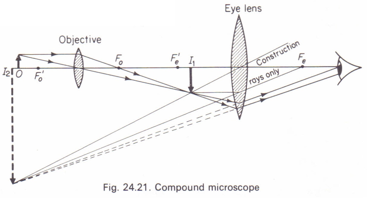
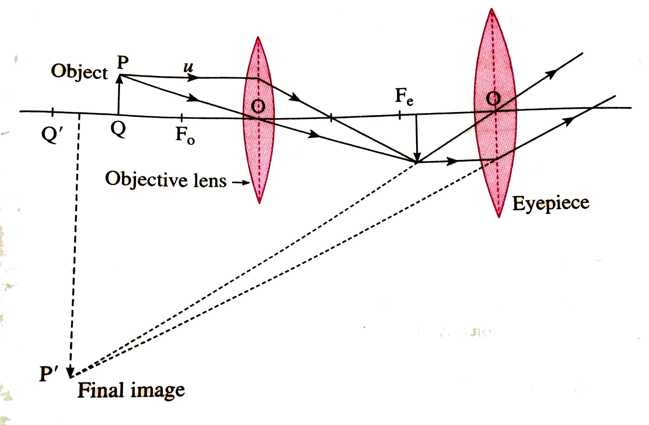
Using a combination of lenses, the working principle of a compound microscope is that a highly magnified image of the specimen is formed at the least possible distance from the distinct vision of an eye that is held very close to the eyepiece of the microscope when the specimen is placed just beyond the focus of the objective lens.

Fixed Focus Microscope Objective Lens Diagram Schematic And Image 02
... principles – image formation, Part 2 of “Understanding the microscope” by Jeremy Sanderson. ... Ray diagrams help us to understand the action of lenses.
What Is An Infinity Corrected Optical System Learn About Microscope Olympus
Multiple lenses and mirrors are used in this microscope. ... A ray diagram from left to right shows a virtual inverted enlarged final image of the.
Anatomy Of A Microscope
Microscope: It is an instrument that produces enlarged images of small objects, allowing the observer an exceedingly close view of minute structures at a scale convenient for examination and analysis.It is scientific equipment that magnifies microscopic objects that are not visible to the naked eyes. Furthermore, with the help of a microscope, we can see various organisms, like a cell, the ...
Solved A Schematic Diagram Of A Refractive Microscope Is Chegg Com
All of the imaging components in the optical microscope are governed by the basic geometrical relationships described above. This includes the collector lens, ...

Understanding Microscopes And Objectives Edmund Optics
Construct the microscope and measure the distance of the object to the objective lens and the distances between the lenses to bring the final image into focus.3 pages
The Microscope An Introduction To Microscopic Methods And To Histology Microscopes 1899 Ch Microscope And Accessories 17 In General The Angle Increases With The Size Of The Lenses Forming The
Microscope Parts and Functions With Labeled Diagram and Functions How does a Compound Microscope Work?. Before exploring microscope parts and functions, you should probably understand that the compound light microscope is more complicated than just a microscope with more than one lens.. First, the purpose of a microscope is to magnify a small object or to magnify the fine details of a larger ...
Microscope Kids Britannica Kids Homework Help
the SPECIMEN, and the LENSES H. DIAPHRAGM Regulates the amount of LIGHT on the specimen E. STAGE Supports the SLIDE being viewed K. ARM Used to SUPPORT the B. NOSEPIECE microscope when carried Holds the HIGH- and LOW- power objective LENSES; can be rotated to change MAGNIFICATION. Power = 10 x 4 = 40 Power = 10 x 10 = 100 Power = 10 x 40 = 400
The Compound Microscope Physics Homework Help Physics Assignments And Projects Help Assignments Tutors Online
Objective Lenses: Usually you will find 3 or 4 objective lenses on a microscope. They almost always consist of 4x, 10x, 40x and 100x powers. When coupled with a 10x (most common) eyepiece lens, total magnification is 40x (4x times 10x), 100x , 400x and 1000x.
Solved Draw A Neat Schematic Diagram Showing The Principle Of A Simple Microscope The Microscope Contains An Objective Lens And An Eyepiece Lens Note 1 Your Diagram Must Include All Information But No
A ray diagram like this can be interpreted in both directions. If you put a point light source one focal length from a converging lens, parallel light ... focal plane of the microscope's objective lens is on the side that faces the specimen. The front focal plane of the condenser is the side that faces the field iris and the illuminator. The ...
Applications Of Mirrors And Lenses
Aperture. Illuminator. Condenser. Diaphragm. Parts of a Compound Microscope. Video: Parts of a compound Microscope with Diagram Explained. As a side note, the microscope used in this post is a great entry level or beginner microscope if you are trying to get someone interested in microscopes, microbiology, or science in general.
Convex Lens Use Microscope
microscope diagram Flashcards. Browse 500 sets of microscope diagram flashcards. Study sets Diagrams Classes Users. 17 Terms. Jeremy-Green. Microscope Diagram. Eyepiece. Nose piece. Coarse Adjustment Knob.
What Is Histology The Histology Guide
A typical compound microscope has two lenses - an objective lens near the specimen and an ocular lens at the top - each of which magnifies the image of the specimen by a certain amount. The ocular lens on most microscopes magnifies 10x (meaning that the image produced by the ocular lens is ten times as large as the specimen).

Ray Diagram Of Focussing On A Compound Microscope Physics Stack Exchange
Labeled part diagram of a stereo microscope Major structural parts of a stereo microscope. There are three major structural parts of a stereo microscope. The viewing Head includes the upper part of the microscope, which houses the most critical optical components, including the eyepiece, objective lens, and light source of the microscope.
Investigations Of The Compound Microscope
The optical microscope, also referred to as a light microscope, is a type of microscope that commonly uses visible light and a system of lenses to generate magnified images of small objects. Optical microscopes are the oldest design of microscope and were possibly invented in their present compound form in the 17th century. Basic optical microscopes can be very simple, although many complex ...
Which Ray Diagram Is Correct For A Compound Microscope Physics Forums
by SJ Ling · 2016 — A compound microscope is composed of two lenses: an objective and an eyepiece. ... Copy and draw rays to find the final image in the following diagram.
Microscopes

Optical Instrument Compound Microscope
Zeiss Microscopy Online Campus Microscopy Basics Numerical Aperture And Resolution
Indian Institute Of Technology
1
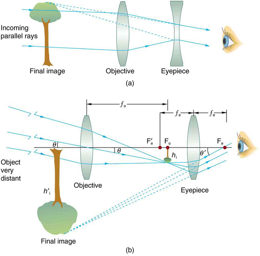
Telescopes Physics
Parts And Components Of Light Microscopes Light Microscope
The Compound Light Microscope

Optical Microscopes Some Basics Science Lab Leica Microsystems

Understanding Microscopes And Objectives Edmund Optics

Infinity Corrected Microscope Building From Scratch Physics Stack Exchange
With A Neat Labelled Diagram Explain The Construction And Work

Optical Microscopes Some Basics Science Lab Leica Microsystems

Light Microscope Vs Electron Microscope Accelerating Microscopy

Draw A Ray Diagram Of A Compound Microscope Diagram Transparent Png 678x317 Free Download On Nicepng
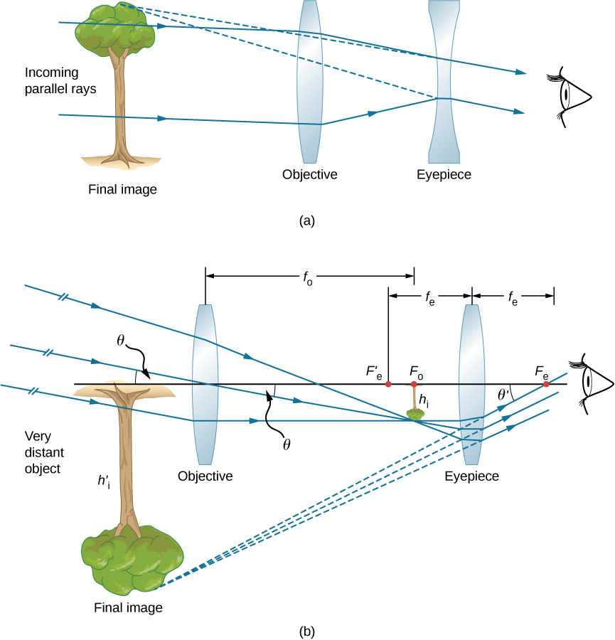
Microscopes And Telescopes University Physics Volume 3

Principles Of Imaging With An Optical Microscope A Ray Diagram Of Download Scientific Diagram
Electricity Detailed Contents


Comments
Post a Comment