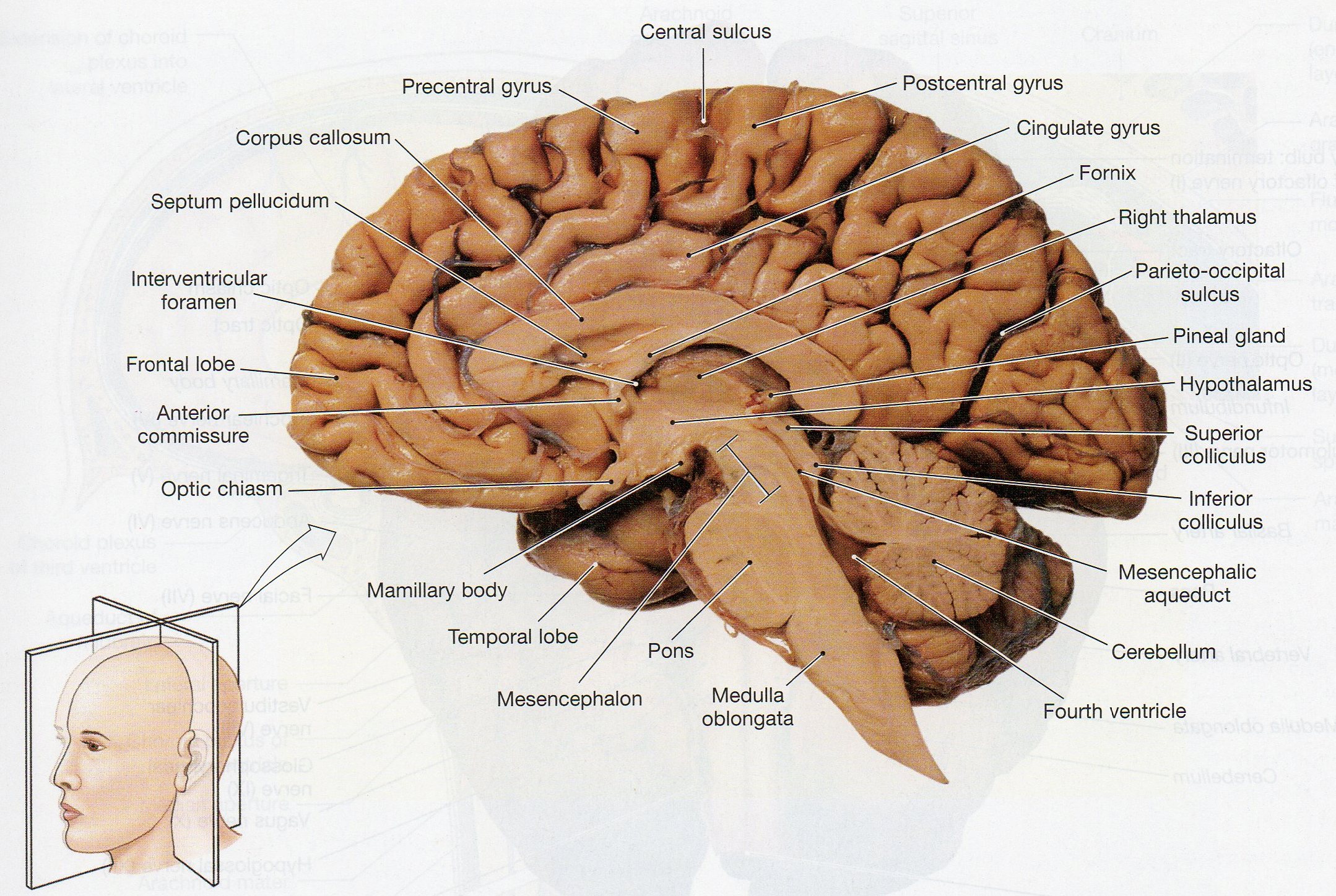
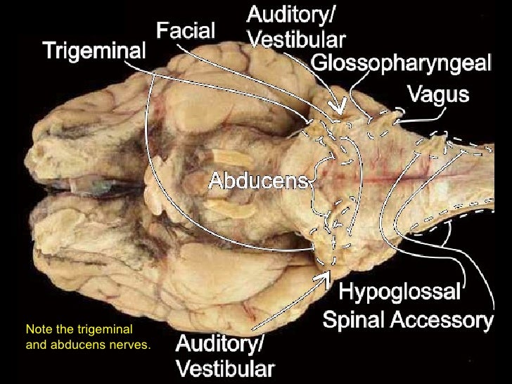
38 sheep brain labeled diagram
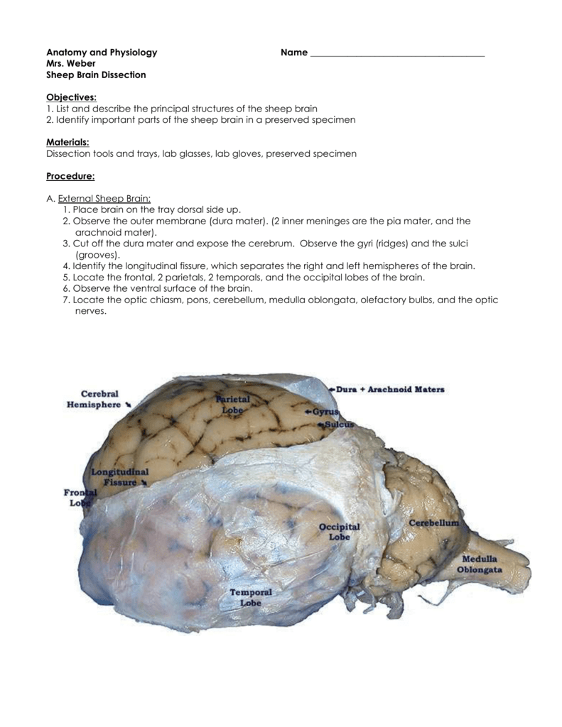
The small intestine begins at the duodenum and is a tubular structure, usually between 6 and 7 m long. Its mucosal area in an adult human is about 30 m 2 (320 sq ft). The combination of the circular folds, the villi, and the microvilli increases the absorptive area of the mucosa about 600-fold, making a total area of about 250 m 2 (2,700 sq ft) for the entire small intestine. The sheep brain is exposed and each of the structures are labeled and described in a sequential manner, in the same way that a real dissection would occur. Sheep Brain Dissection. 1. The sheep brain is enclosed in a tough outer covering called the dura mater. You can still see some structures on the brain before you remove the dura mater.
Start studying Sheep Brain Dissection labeled. Learn vocabulary, terms, and more with flashcards, games, and other study tools.

Sheep brain labeled diagram
Sheep Brain Dissection with Labeled Images The sheep brain is exposed and each of the structures are labeled and described in a sequential manner, in the same way that a real dissection would occur. A Amanda Huss Anatomy Brain Science Medical Science Medical Coding Nursing School Notes Medical School Ob Nursing Nursing Schools Brain Diagram Diagram Worksheets. Label the Parts of a Sheep Brain. Print out these diagrams and fill in the labels to test your knowledge of sheep brain anatomy. Internal anatomy: label the right side (.pdf) External anatomy: label the top view (.pdf) External anatomy: label the bottom view (.pdf) What other users say: Fun and Educational. The brain is contained within the cranial cavity of the skull, and the spinal cord is contained within the vertebral cavity of the vertebral column. It is a bit of an oversimplification to say that the CNS is what is inside these two cavities and the peripheral nervous system …
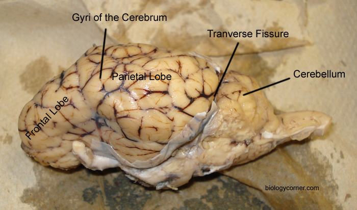
Sheep brain labeled diagram. An extremely efficient memory center in the brain. Responsible for spatial memory and spatial navigation as well as some types of non-spatial memory such as contextual memory and episodic memory. Damage to this structure produces both anterograde and retrograde amnesia. - Vertical cut through pineal gland laterla to pineal gland. Confocal analysis of the labeled tumors demonstrated that Dil-liposomes specifically labeled a population of pvTAMs (Fig. 2, J to M, and fig. S4, A to C) and ex vivo characterization of the Dil-labeled TAMs in enzyme-dispersed MMTV-PyMT tumors confirmed the vast majority of labeled cells to be that of the Lyve-1 + TAM subset . Methods and Materials: We will be dissecting a preserved adult sheep brain. Our dissection kit contains a scalpel, a fine tipped pair of scissors, a blunt metal probe, a fine tipped forceps, a blunt-tipped forceps. It is vital to wear vinyl gloves. It is important to be careful when working with preservatives. Sheep Neuroanatomy Lab- Labeling Worksheet Psychology 2315- Brain and Behaviour Kwantlen Polytechnic University Figure 1: Dorsal view Cerebellum, Frontal lobe, Occipital lobe, Parietal lobe, and Temporal lobe. Temporal Parietal Lobe Frontal Lobe Cerebellum Occipital Lobe
5 3 11 6 22 16 18 1. Gray Matter 2. White Matter 3. Corpus Callosum 4. Lateral Ventricle 5. Caudate Nucleus 6. Septum Pellucidum 7. Fornix 8. Take A Sneak Peak At The Movies Coming Out This Week (8/12) ‘Not Going Quietly:’ Nicholas Bruckman On Using Art For Social Change; New Movie Releases This Weekend: December 10-12 Molecular Biology, Robert Weaver, 5th Edition BI 335 - Advanced Human Anatomy and Physiology Western Oregon University Figure 4: Mid-sagittal section of brain showing diencephalon (includes corpus callosum, fornix, and anterior commissure) Marieb & Hoehn (Human Anatomy and Physiology, 9th ed.) - Figure 12.10 Exercise 2: Utilize the model of the human brain to locate the following structures / landmarks for the
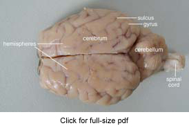
The primary function of the meninges and of the cerebrospinal fluid is to protect the central nervous system. Dura Mater-encases the prain and is the first layer of the brain. Gyrus - a ridge or fold between two clefts on the cerebral surface in the brain. Sulcus - a groove or furrow, especially one on the surface of the brain. May 28, 2015 · Electron microscope is also employed to detect tissue cysts in mouse brain and oocysts in the small intestine of infected cats, but it is difficult to be applicable for routine use [29, 30]. Bioassay The isolation of T. gondii by bioassay using laboratory animals is generally considered as the gold standard for detection of T. gondii infection. Sheep Brain Neuroanatomy Online Self-Test. Use each diagram as a reference, and selected the correct answer for each lettered structure. You may find it useful to open the diagrams in a separate window to review while answering each question. Dorsal Surface. This is an online quiz called sheep brain labeling. There is a printable worksheet available for download here so you can take the quiz with pen and paper. Your Skills & Rank. Total Points. 0. Get started! Today's Rank--0. Today 's Points. One of us! Game Points. 22. You need to get 100% to score the 22 points available.
Start studying Sheep Brain Dissection labeled 2. Learn vocabulary, terms, and more with flashcards, games, and other study tools.
DISSECTION OF THE SHEEP'S BRAIN Introduction The purpose of the sheep brain dissection is to familiarize you with the three-dimensional structure of the brain and teach you one of the great methods of studying the brain: looking at its structure. One of the great truths of studying biology is the saying that "anatomy precedes physiology".
Sheep are wonderful and cute. The brain is an interesting organ. It helps with cognition and memory. Almost all the basic task In the body is commanded by the Brain. It is the control center of the body which regulates and control the process crucial for survival Are you interested in learning more about the brain of different animals? Can you answer all the questions of this "Sheep Brain ...
to anatomy studies. See for yourself what the . cerebrum, cerebellum, spinal cord, gray matter, white matter, and other parts of the brain look like! Observation: External Anatomy . 1. You'll need a . preserved sheep brain. for the dissection. Set the brain down so the flatter side, with the white . spinal cord. at one end, rests on the ...
Shows pictures of a sheep and a human brain. Each of the 12 cranial nerves is represented, students color and number each nerve in both brains. Learn the external and internal anatomy of sheep brains with HST's Learning Center science lesson and guide! Diagram worksheets also included.
Image Result For Sheep Brain Labeled Brain Diagram Human Brain Diagram Brain Anatomy. Sheep Brain Dissection Project Guide Hst Learning Center Dissection Brain Mapping Science Biology. Sheep Brain Dissection Lab Companion In 2021 Brain Anatomy Anatomy And Physiology Brain. Sheep Brain External View Labeled Anatomia Veterinaria Anatomia Veterinaria.
Aug 04, 2015 · A, Diagram of the mouse placenta illustrating the 3 different regions: 1) maternal decidua, 2) junctional zone, and 3) labyrinithine zone. In the labyrinthine region, fetal blood is separated from maternal blood by 3 trophoblast layers: 2 synctiotrophoblast layers and 1 cytotrophoblast layer, which directly contacts the maternal blood, giving ...
This map shows the major structures of the sheep brain with an active cursor to help identify the structures. Neuroanatomy Tutorial I: Basic Anatomy of the Brain. Point to any region of this midsaggital sheep brain image (medial view) to highlight that structure. Click the left mouse button to identify the structure you are pointing to.
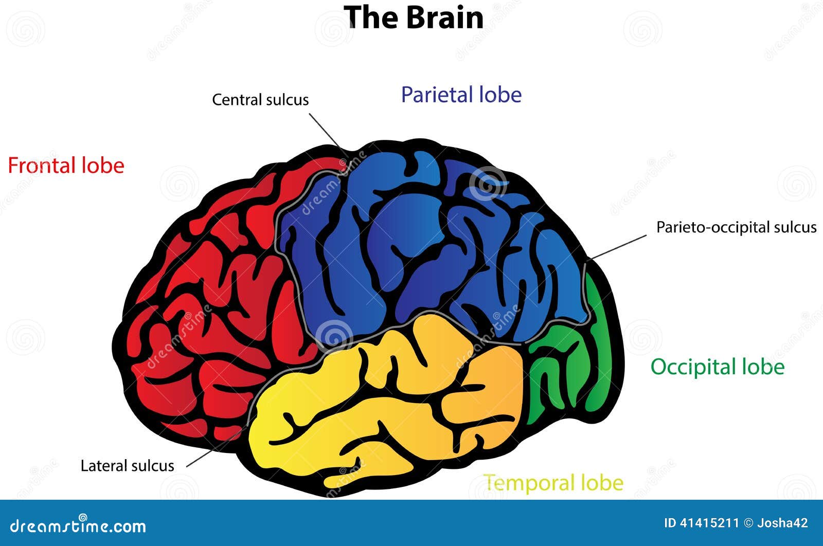
2,683 labeled brain anatomy stock photos, vectors, and illustrations are available royalty-free. See labeled brain anatomy stock video clips. of 27. brain diagram with labels hypothalamus vector brain diagram pons cerebrum and cerebellum brain pons brain anatomy amygdala brain labelled amygdala brain human midbrain diagram pons.
Use this printable frog dissection diagram with labeled parts (.pdf) as a guide for locating them. Heart. The frog’s heart is the small triangular organ at the top. Unlike a mammal heart, it only has three chambers — two atria at the top and one ventricle below. Carefully cut away the pericardium, the thin membrane surrounding the heart.
The male gonad is the testis (pl, testes).. The initial difference in male and female gonad development are dependent on testis-determining factor (TDF) the protein product of the Y chromosome SRY gene. Recent studies have indicated that additional factors may also be required for full differentiation.
Sheep Brain Dissection Labeled Diagram. angelo. October 16, 2021. Image Result For Sheep Brain Labeled Brain Diagram Human Brain Diagram Brain Anatomy. Sheep Brain External View Labeled Anatomia Veterinaria Anatomia Veterinaria. Pin By Aleena Hanson On A P Brain Anatomy Anatomy Brain Images. Mit Hhmi Summer Workshop For Teachers Neuroanatomy ...
Anatomy and Physiology. Anatomy and Physiology questions and answers. Art-labeling Activity: Midsagittal Section of the Sheep Brain (Diagram, 2 of 2) Reset Help Fomix Infundibulum Olfactory bulb Optic chiasm Mosencephalon Pituitary gland Marillary body Medulla oblongata Pons Spinal cord Corpus callosum Art-labeling Activity: Midsagittal Section ...
Why$dread$a$bump$on$the$head?$ $ October$2012$ Lesson$2:$What$does$thebrain$looklike?$ $ $ $ 2 $ What'supwithallthenewwords?! $ With$all$of$the$new$terminology$in ...
301 Moved Permanently. nginx
The lobes of the brain are visible, as well as the transverse fissure, which separates the cerebrum from the cerebellum. The convolutions of the brain are also visible as bumps (gyri) and grooves (sulci). Use the diagram below to help you locate these items. Dorsal View of the Sheep Brain . 8.
function, and pathology. Those students participating in Sheep Brain Dissections will have the opportunity to dissect and compare anatomical structures. At the end of this document, you will find anatomical diagrams, vocabulary review, and pre/post tests for your students. The following topics will be covered: 1.
The clitoris (/ ˈ k l ɪ t ər ɪ s / or / k l ɪ ˈ t ɔːr ɪ s / ()) is a female sex organ present in mammals, ostriches and a limited number of other animals.In humans, the visible portion – the glans – is at the front junction of the labia minora (inner lips), above the opening of the urethra.
Start studying sheep brain (half). Learn vocabulary, terms, and more with flashcards, games, and other study tools.
Sheep Brain Anatomy Lab Manual. Based on original material by R. N. Leaton, Dartmouth College. Contributors to this version: Al Sorenson, Lisa Raskin, Sarah Turgeon, Steve George, and JP Baird. I. Introduction. The brain of the sheep is useful for study because its anatomy is similar to human brain anatomy. Although exact proportions (and names ...
Your brain on movies answer key quizlet. 0 Cart. Your brain on movies answer key quizlet ...
Oct 07, 2020 · 48 Likes, 2 Comments - College of Medicine & Science (@mayocliniccollege) on Instagram: “🚨 Our Ph.D. Program within @mayoclinicgradschool is currently accepting applications! As a student,…”
Professional academic writers. Our global writing staff includes experienced ENL & ESL academic writers in a variety of disciplines. This lets us find the most appropriate writer for any type of assignment.
Index Of Files Occ Video Upload Faculty Resources Acamilo Academics Biol 130l Practical 2 Sheep Brain 1
The sheep brain is quite similar to the human brain except for proportion. The sheep has a smaller cerebrum. Also the sheep brain is oriented anterior to posterior whereas the human brain is superior to inferior. 1. The tough outer covering of the sheep brain is the dura mater, one of three meninges (membranes) that cover the brain. You will ...
The brain is contained within the cranial cavity of the skull, and the spinal cord is contained within the vertebral cavity of the vertebral column. It is a bit of an oversimplification to say that the CNS is what is inside these two cavities and the peripheral nervous system …
Diagram Worksheets. Label the Parts of a Sheep Brain. Print out these diagrams and fill in the labels to test your knowledge of sheep brain anatomy. Internal anatomy: label the right side (.pdf) External anatomy: label the top view (.pdf) External anatomy: label the bottom view (.pdf) What other users say: Fun and Educational.
Sheep Brain Dissection with Labeled Images The sheep brain is exposed and each of the structures are labeled and described in a sequential manner, in the same way that a real dissection would occur. A Amanda Huss Anatomy Brain Science Medical Science Medical Coding Nursing School Notes Medical School Ob Nursing Nursing Schools Brain Diagram










Comments
Post a Comment