43 guard cells diagram
Jun 25, 2007 · Guard cells use osmotic pressure to open and close stomata, allowing plants to regulate the amount of water and solutes within them. . Critical in this process is the stoma. Stomata (multiple stoma) are located on the outermost cellular layer of leaves, stems, and other plant parts. An open stoma facilitates the process of photosynthesis in ... 7 The diagram shows a cross-section through a plant stem. Q Q shows the part that is stained red when the stem is placed in water containing a red dye. What is found at Q? A guard cells B palisade cells C phloem D xylem 8 The diagram shows a motor (effector) neurone. Which structure is also found in white blood cells, but not in red blood cells ...
in cells P, Q and R. Diagram 2 ... Guard cells Phloem cells Xylem cells (1) (ii)€€€€€Describe how the appearance of the stomata in leaf B is different from the appearance of the stomata in leaf A. _____ _____ (1) Page 18 of 19 www.examqa.com (iii)€€€€The man forgets to water the plant. What might happen to the plant in the ...

Guard cells diagram
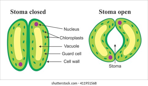
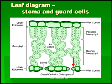
Expert Answer: The opening and closing of stomata are controlled by the guard cells. When water flows into the guard cells, they swell up and the curved surface causes the stomata to open. When the guard cells lose water, they shrink and become flaccid and straight thus closing the stomata. The cell wall surrounding the pore is tough and flexible. The shape of guard cells usually differs in both monocots and dicots, though the mechanism continues to be the same. Guard cells are bean-shaped and contain chloroplasts. They contain chlorophyll and capture light energy. The subsidiary cells surround the guard cells. ei the following parts of the leaf in the diagram below. Give the purpose/function of lower epidermis upper epidermis patisade layer cuticle stomate guard cells vein (fibrovascular bundle) spongy layer air space xylem phloem chloroplasts mesophyll Onstructional Pct , F8765 a. b h. Name Stomata Class Date The stomata of a plant open and close to ...
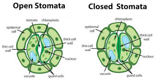
Guard cells diagram. The diagram below shows the direction of movement of substances through a plant. ... guard cells palisade cells mesophyll cells (1) (b) Water loss by evaporation from leaves is called transpiration. A student set up an experiment to investigate water loss from leaves. The student: • took ... Thus, the guard cells swell. The walls of guard cells of stomatal pore are thicker outside and the walls present inside are thinner, guard cells bulge to due to the inflow of water, thus widening the stomatal opening. When the osmotic pressure of the guard cells becomes lower (during the night), the water leaves these cells due to exosmosis and ... Guard cells are cells surrounding each stoma. They help to regulate the rate of transpiration by opening and closing the stomata. Light is the main trigger for the opening or closing. Each guard cell has a relatively thick cuticle on the pore-side and a thin one opposite it. The diagram represents specialized cells in the surface of the leaf of a green plant. The main function of these cells is to. ... The diagram below represents a change in guard cells that open and close pores in a plant. This change directly helps to. answer choices . increase heterotrophic nutrition.
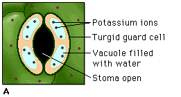
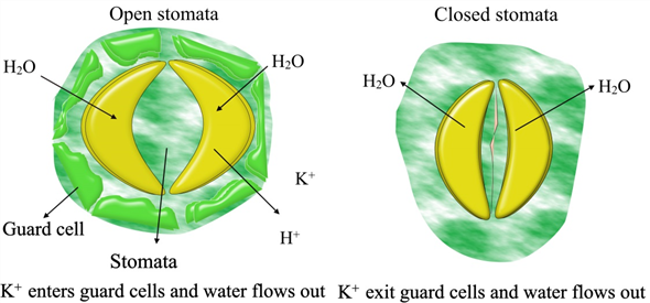
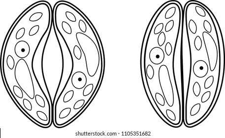
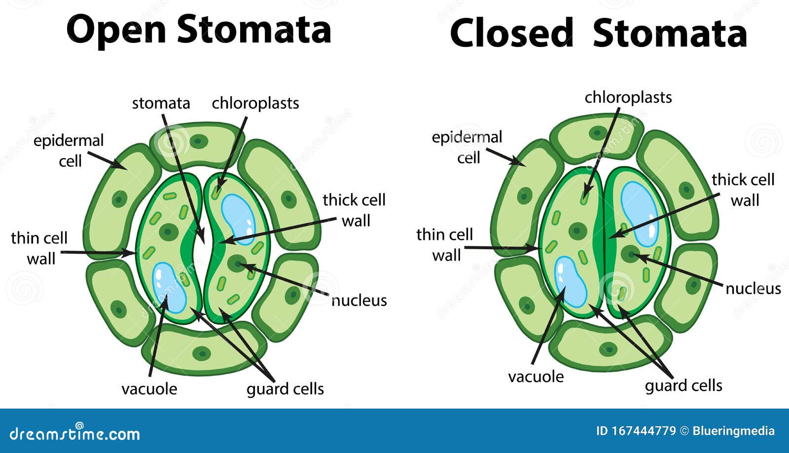
Answer (1 of 2): Leaves have special pores called stomata that make gas exchange possible while helping to control the loss of water. The stomata operate through the use of two tiny jellybean shaped cells called guard cells located in the outer layer of tissue called the epidermal layer. Guard ce... Essentially, guard cells are two bean-shaped cells that surround a stoma. As epidermal cells, they play an important role in gaseous exchange in and out of plant leaves by regulating the opening and closing of pores known as a stoma. In addition, they are the channels through which water is released from leaves to the environment. As such, guard cells play a crucial role in photosynthesis by regulating the entry of materials necessary for the process. Apart from regulating gaseous exchange (as well as water release from leaves), they have also been shown to contain chloroplasts which also make them a site of photosynthesis. Some of the factors that influence guard cell activities include: 1. Humidity 2. Temperature 3. Light 4. Carbon dioxide 5. Potassium ions 6. Hormones *In Greek, the word "stoma" means mouth. *Although stomata are commonly found in plant leaves, they can also be found in the stems. (e)€€€€€The diagram below shows two guard cells surrounding a closed stoma and two guard cells surrounding an open stoma. € When light intensity is high potassium ions are moved into the guard cells. Describe how the movement of potassium ions into the guard cells causes the stoma to open. Leaf anatomy vector illustration diagram. Biological macro scheme poster. Leaf anatomy vector illustration diagram. Biological macro scheme poster with leaf inner layers, veins and breathing oxygen exchange. guard cells stock illustrations
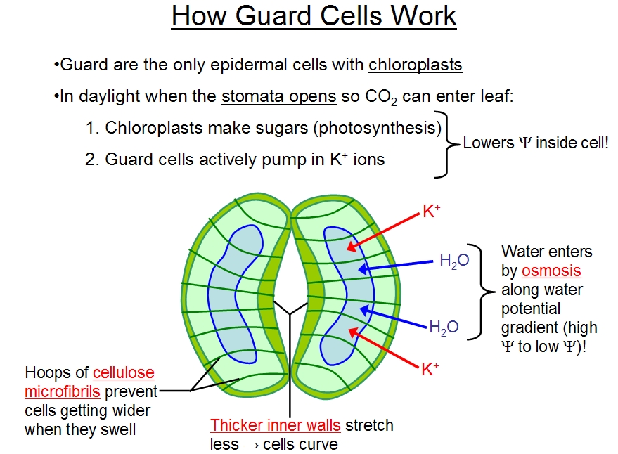
Guard Cells These cells resemble the shape of a kidney or dumbbell-shaped that consists of the chloroplast. They contain chlorophyll and capture light energy. The primary function of guard cells is to properly carry out the opening and closing mechanism of the stoma. Guard cells have numerous ectodesmata.The guard cells become turgid when the water concentration is high within the cell than the surrounding. Conversely, the low concentration of water within the cell than the surrounding makes the guard cells flaccid.. Organelles within the guard cells: Microtubules orient cellulose microfibrils and contribute to building guard cells. Create a labeled diagram that explains how guard cells sense blue light and respond by becoming turgid. Include PHOT, H+ pump, hyperpolarization-gated K+ channels, H+/solute cotransporters, solute concentration, H2O, and pressure potential. B-represents guard cells. kigney shaped and dumbel shaped respectively. C-represent subsidiary cells - epidermal cells surrounding the guard cells, maintain turgor pressure of the guard cells. So, the correct answer is ' A-Stoma ,B-Guard cells,C- Subsidiary cells'
Characteristics Of Guard Cell Chloroplasts And Their Possible Functions In Stomata Behavior Plant Stomata Encyclopedia
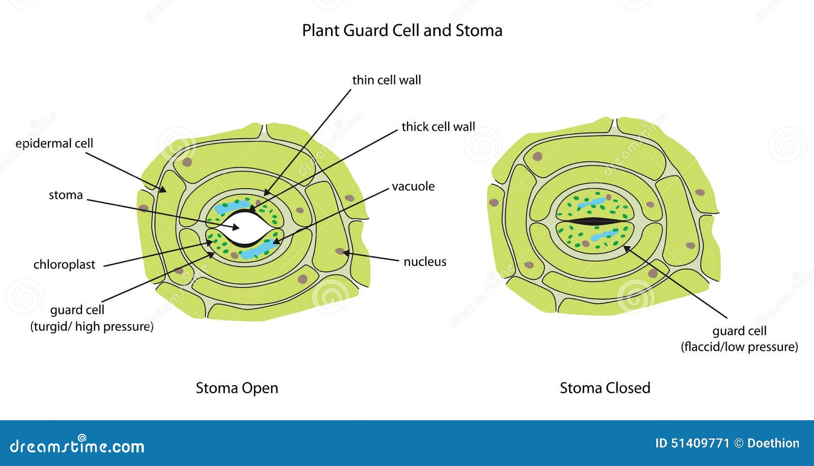
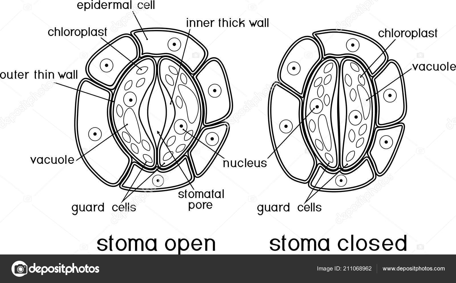
The guard cells are bean-shaped in dicots and dumb-bell shaped in monocots. The inner wall of the guard cell is thick whereas the outer wall is thin. Plants need gases like oxygen for respiration and carbon dioxide for photosynthesis. It is through this small opening called stomata the gases diffuse inside the leaf and are taken in.

Guard Cell Wikipedia
How Guard Cells Work • To open: • Water flows into the guard cells, pushing out on the cell membrane rings prevent increase of cell in diameter cell increases in length but outer wall is more flexible and bulges outwards inner wall is pulled and stoma opens
Transpiration How Do Guard Cells Function
Jun 18, 2021 · Guard cells are a pair of bean-shaped cells found in the epidermis of leaves and young stems of plants. They look similar to a kidney and exist in pairs surrounding a tiny gas exchange opening called a stoma. Guard cells help plants to perform photosynthesis, get rid of wastes, and excess water. Guard Cells Diagram.

Stomata Definition Diagrams And Functions
Aug 31, 2021 · The guard cells change shape depending on the amount of water and potassium ions present in the cells themselves. When the guard cells take in potassium ions, water diffuses into the cells by osmosis.

Guard Cells In Plants What Are Guard Cells Video Lesson Transcript Study Com
(2) only guard cells (3) guard cells and epidermal cells (4) guard cells, epidermal cells and stoma Answer: Only guard cells and epidermal cells will be visible under the high power of a microscope. Hence, the correct answer is option 3. Question 5. Which structure out of I, II, III and IV marked in the given diagram of the epidermal peel of ...

Learn About Guard Cell Chegg Com
Lower Epidermis: A protective layer of cells. The lower epidermis produces a waxy cuticle too in some plant species. The lower epidermis contains pores called stomata that allow carbon dioxide and oxygen to move in and out of the plant respectively. Stomata: Tiny pores (small holes) surrounded by a pair of sausage shaped guard cells.

A Draw A Diagram To Show Open Stomatal Pore And Label On It 1 Guard Cells 2 Chloroplast B State Brainly In
Solution. Verified by Toppr. Correct option is C) The above figure shows the complex structure of the stomatal apparatus which consists of a stoma, two bean-shaped guard cells and surrounding 4-5 subsidiary (epidermal) cells. From the question figure, it is clear that label 1,2,3 represents guard cells, stoma and epidermal cells respectively.
Diagram Showing Stomata And Guard Cell Diagram Illustration Stock Vector Image Art Alamy
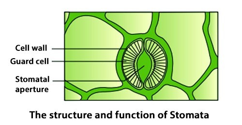
The epidermal cells bordering the guard cells are called accessory cells or subsidiary cells. Generally the term stoma is applied to the stomatal opening and the guard cells. The guard cells are living and contain chloroplasts in them. They also contain a larger proportion of protoplasm than other epidermal cells.

Types Of Stomata In Plants
The stomatal pore is surrounded by two specialized epidermal cells which are known as guard cells. These guard cells are kidney shaped in dicot and dumbel shaped in monocots. The epidermal cells surrounding the guard cells are specialized and are known as subsidiary cells or accessory cells.
Free Vector Diagram Showing Stomata And Guard Cell Diagram
Active transport of water out of the guard cells b. Decreased turgor pressure in guard cells ... The diagram below shows a cross section through a leaf. Drag the labels to the appropriate targets to match the function with the structure indicated in the diagram. Labels may be used once, more than once, or not at all.
Plant Guard Cells With Stoma Fully Labeled Stock Illustration Illustration Of Epidermal Stomata 51409771
Guard Cells: The Guard cell has the shape of a bean or kidney and may also be dumbbell-shaped. The function of the stomata is the gaseous exchange. Hence there is a mechanism of opening and closing. The guard cells control this mechanism. They also maintain the transpiration rate to prevent excessive water loss.
Guard Cells Images Stock Photos Vectors Shutterstock
The diagram below represents specialized cells in the surface of the leaf of a green plant.The main function of these cells is to answer choices change the size of the stomate to regulate water loss close the stomate to keep dust and dirt out of the leaf directly provide leaf cells with the water involved in photosynthesis

Transport Of Materials In A Flowering Plant Stoma Biology Diagrams Cell Diagram
There are present numerous stomata in this layer. Each stoma is surrounded by two guard cells and is followed by a substomatal cavity. Such type of leaf having stomata only on the lower side are known as hypostomatic leaf. The guard cells of stomata may be situated either in the level of epidermal cells or can be placed in pits. 2. Isobilateral ...
Solved What Are The Functions Of Stomata And Lenticels In What W Chegg Com
3 Fig. 8.1 shows a diagram of a stoma, its guard cells and adjacent epidermal cells. epidermal cell guard cells nucleus cytoplasm stoma nucleus. Fig. 8.1 (a) Guard cells have chloroplasts while epidermal cells do not have chloroplasts. State . one other. difference, visible in Fig. 8.1, between guard cells and epidermal cells.
Stomata Structure Guard Cells Biological Science Picture Directory Pulpbits Net
(e)€€€€€The diagram below shows two guard cells surrounding a closed stoma and two guard cells surrounding an open stoma. € When light intensity is high potassium ions are moved into the guard cells. Describe how the movement of potassium ions into the guard cells causes the stoma to open.
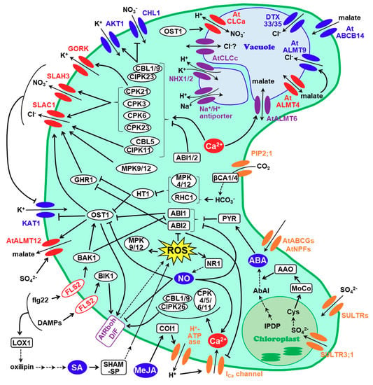
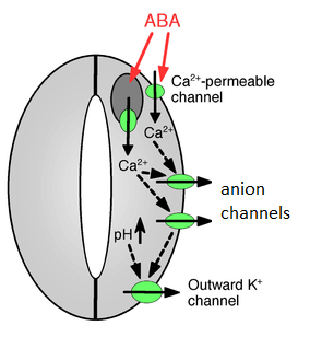
Plants Free Full Text Guard Cell Membrane Anion Transport Systems And Their Regulatory Components An Elaborate Mechanism Controlling Stress Induced Stomatal Closure
Guard cells line the openings of stoma and other organs in plants, opening and closing to moderate the process of respiration. The chief role of guard cells is to prevent an excess loss of water through respiration, allowing the plant to trade oxygen and carbon dioxide without becoming dehydrated.
Stomata Royalty Free Stomata Vector Images Drawings Depositphotos
The diagram shows a cross-section through a plant stem. Q shows the part that is stained red when the stem is placed in water containing a red dye. What is found at Q? A. Guard cells. B. Palisade cells. C. Phloem. D. Xylem. Medium. Open in App. Solution. Verified by Toppr. Correct option is . D. Xylem.

How To Draw The Structure Of Guard Cell Stomata Guard Cell Labelled Diagram Abhishek Educare Youtube
Guard cells: They are the kidney-shaped or dumbbell-shaped cell, which functions by controlling the mechanism (opening and closing) of stomata. This was a brief introduction of the Diagram of Stomata. For more information about Stomata, its structure, functions and other related topics, visit us at BYJU'S Biology.

How Do Guard Cells Of A Leaf Help To Maintain Homeostasis In A Plant Socratic
ei the following parts of the leaf in the diagram below. Give the purpose/function of lower epidermis upper epidermis patisade layer cuticle stomate guard cells vein (fibrovascular bundle) spongy layer air space xylem phloem chloroplasts mesophyll Onstructional Pct , F8765 a. b h. Name Stomata Class Date The stomata of a plant open and close to ...

Stomatal Opening Osmosis Questions From Test Diagram Quizlet
The cell wall surrounding the pore is tough and flexible. The shape of guard cells usually differs in both monocots and dicots, though the mechanism continues to be the same. Guard cells are bean-shaped and contain chloroplasts. They contain chlorophyll and capture light energy. The subsidiary cells surround the guard cells.

Diagram Showing Stomata And Guard Cell Illustration Canstock
Expert Answer: The opening and closing of stomata are controlled by the guard cells. When water flows into the guard cells, they swell up and the curved surface causes the stomata to open. When the guard cells lose water, they shrink and become flaccid and straight thus closing the stomata.

Stomata Royalty Free Stomata Vector Images Drawings Depositphotos
Guard Cell Images Stock Photos Vectors Shutterstock
17 1 2 2 Stomatal Opening And Closure Biology Libretexts
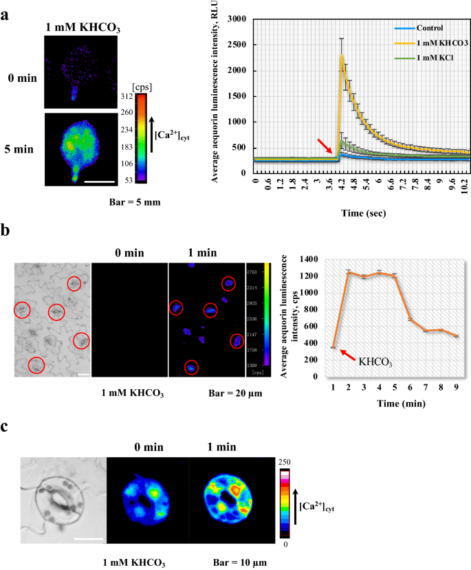
Arabidopsis Guard Cell Co2 Hco3 Response Mutant Screening By An Aequorin Based Calcium Imaging System Plant Methods Full Text
Vajorna 1738 G Stomata All Viucus Lupppiiiy I Og Gr 4 Diagram Microscope Stomata Guard Cells
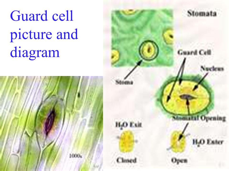
Guard Cells By Shelby Stutzman Guard Cell Picture And Diagram Ppt Download
Explain The Structure Of Stomata Write Function Of Guard Cells Snapsolve

Plant Guard Cells With Stoma Fully Labeled Canvas Print Barewalls Posters Prints Bwc25963385
Plantae Review Guard Cell Metabolism And Stomatal Function Annu Rev Plant Biol Plantae

Vector Stock Plant Guard Cells With Stoma Fully Labeled Clipart Illustration Gg76570934 Gograph
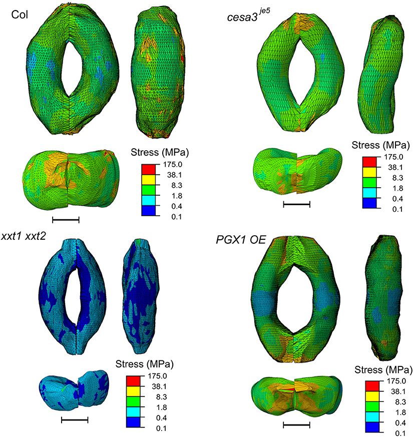
Frontiers Mechanical Effects Of Cellulose Xyloglucan And Pectins On Stomatal Guard Cells Of Arabidopsis Thaliana Plant Science
Guard
Open Stomata Stock Illustrations 25 Open Stomata Stock Illustrations Vectors Clipart Dreamstime
Plants General Structure Presentation Plants Animals And Ecosystems

A What Are Guard Cells Explain Their Role In Regulati Wiredfaculty

Open Stomata Stock Illustrations 25 Open Stomata Stock Illustrations Vectors Clipart Dreamstime
1

Diagram Showing Leaf Guard Cells On Isolated Vector Image

Guard Cell Diagram Diagram Quizlet

Plant Guard Cells Vector Photo Free Trial Bigstock

Diagram Showing Stomata And Guard Cell Royalty Free Vector

Guard Cell Photosynthesis And Stomatal Function Lawson 2009 New Phytologist Wiley Online Library


Comments
Post a Comment