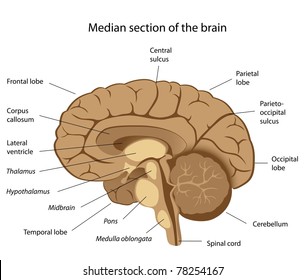
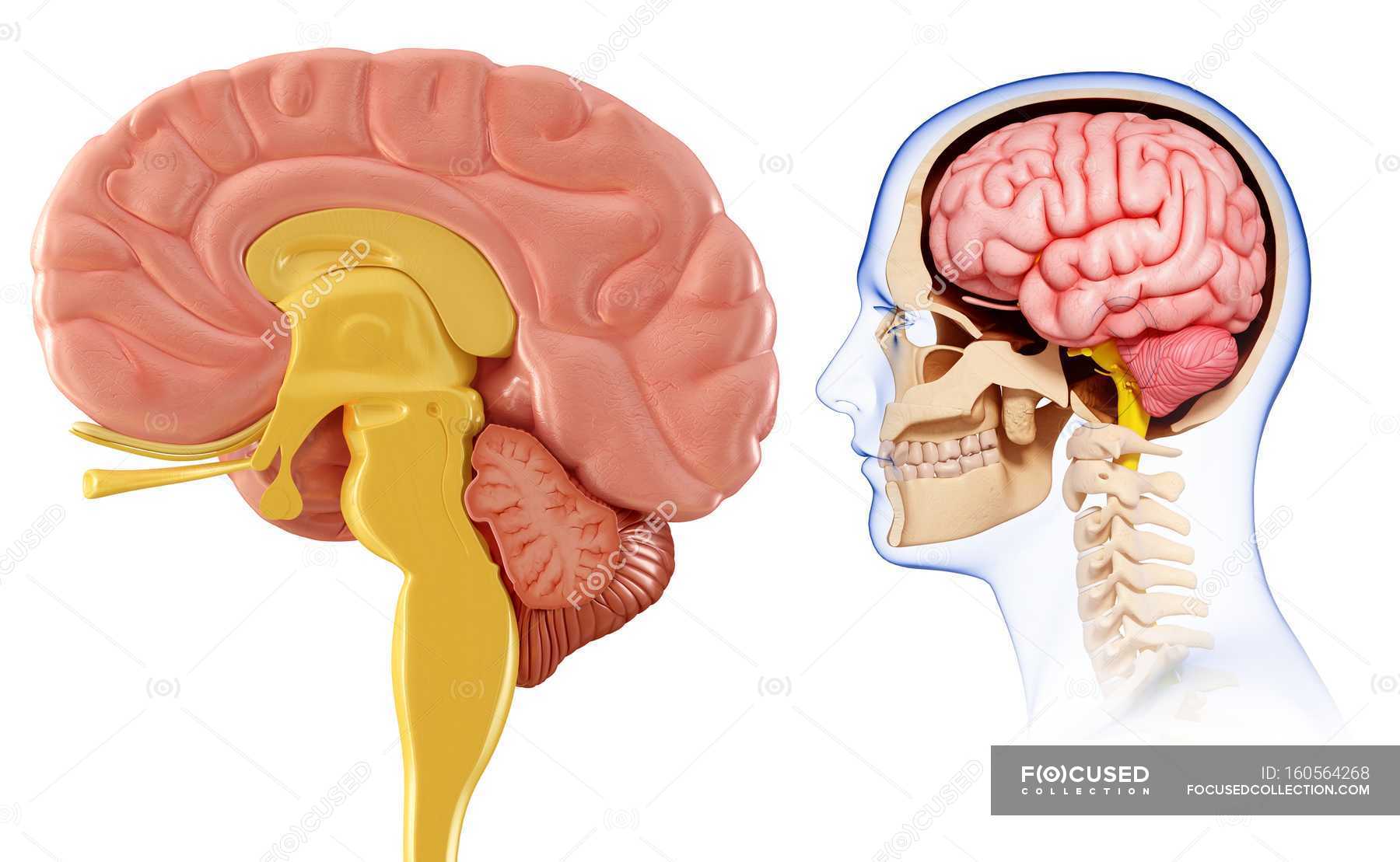
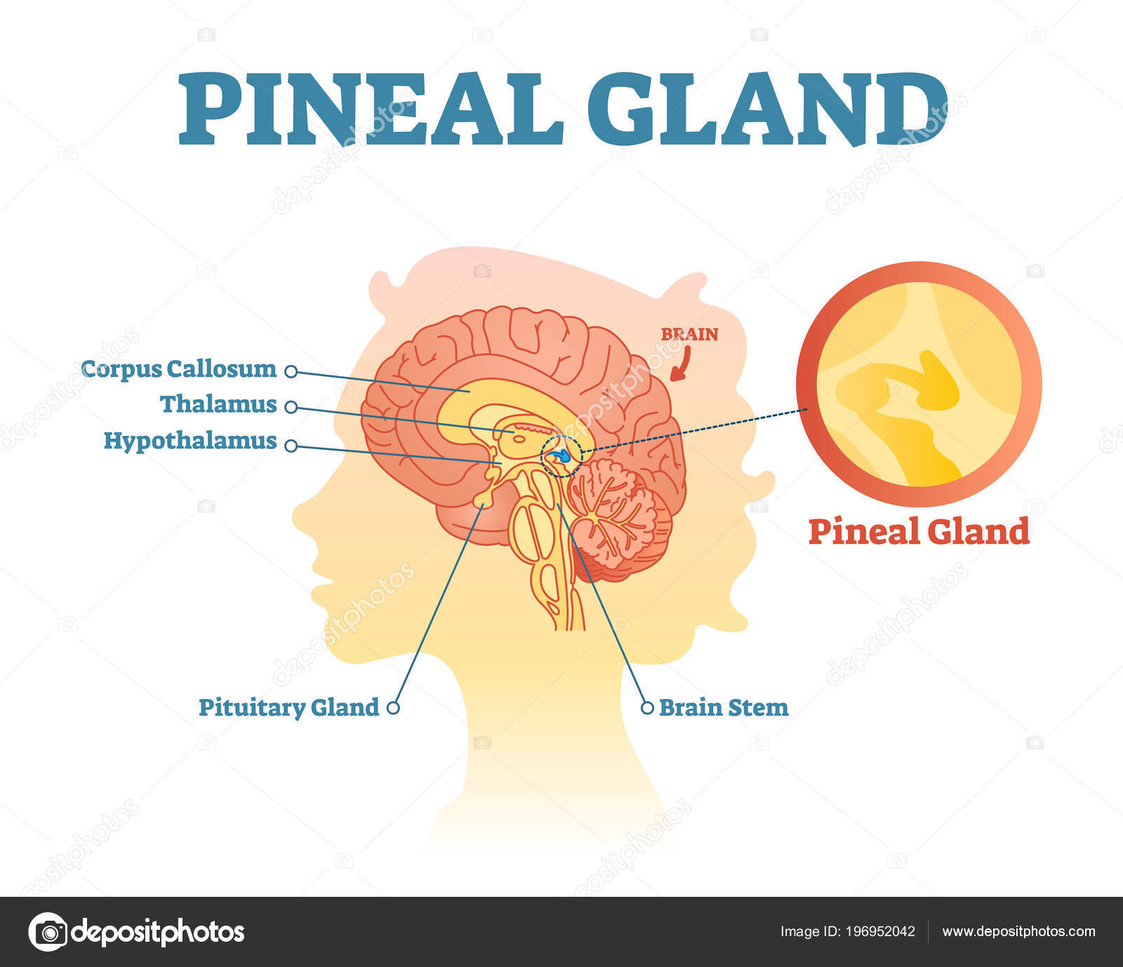
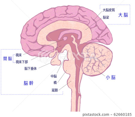
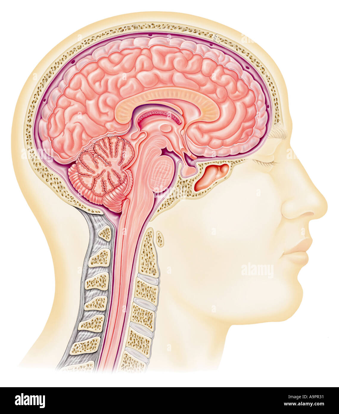
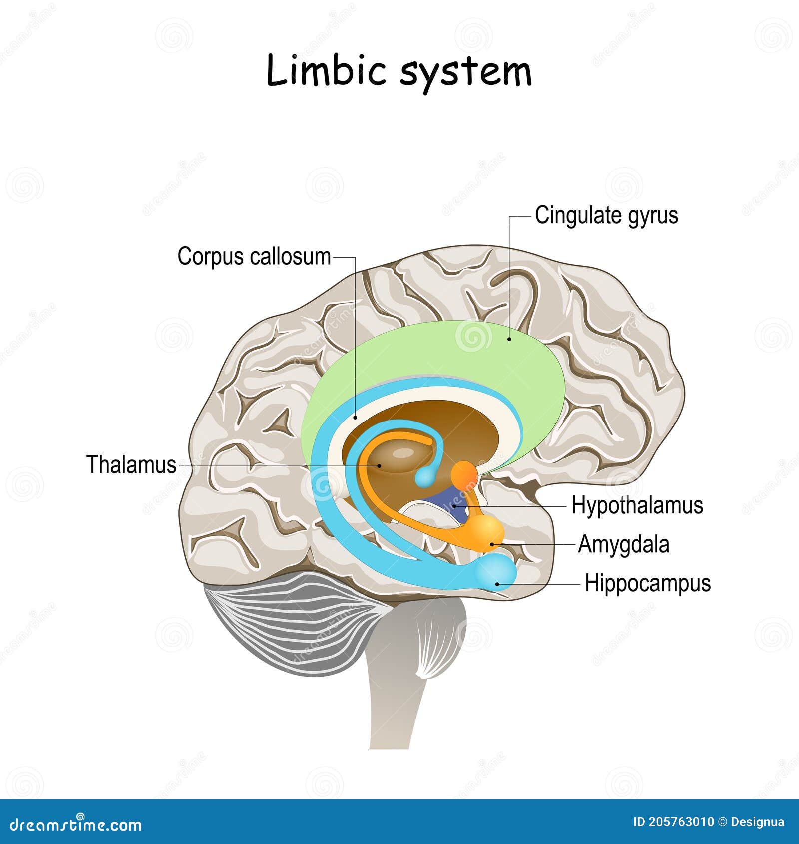
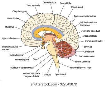
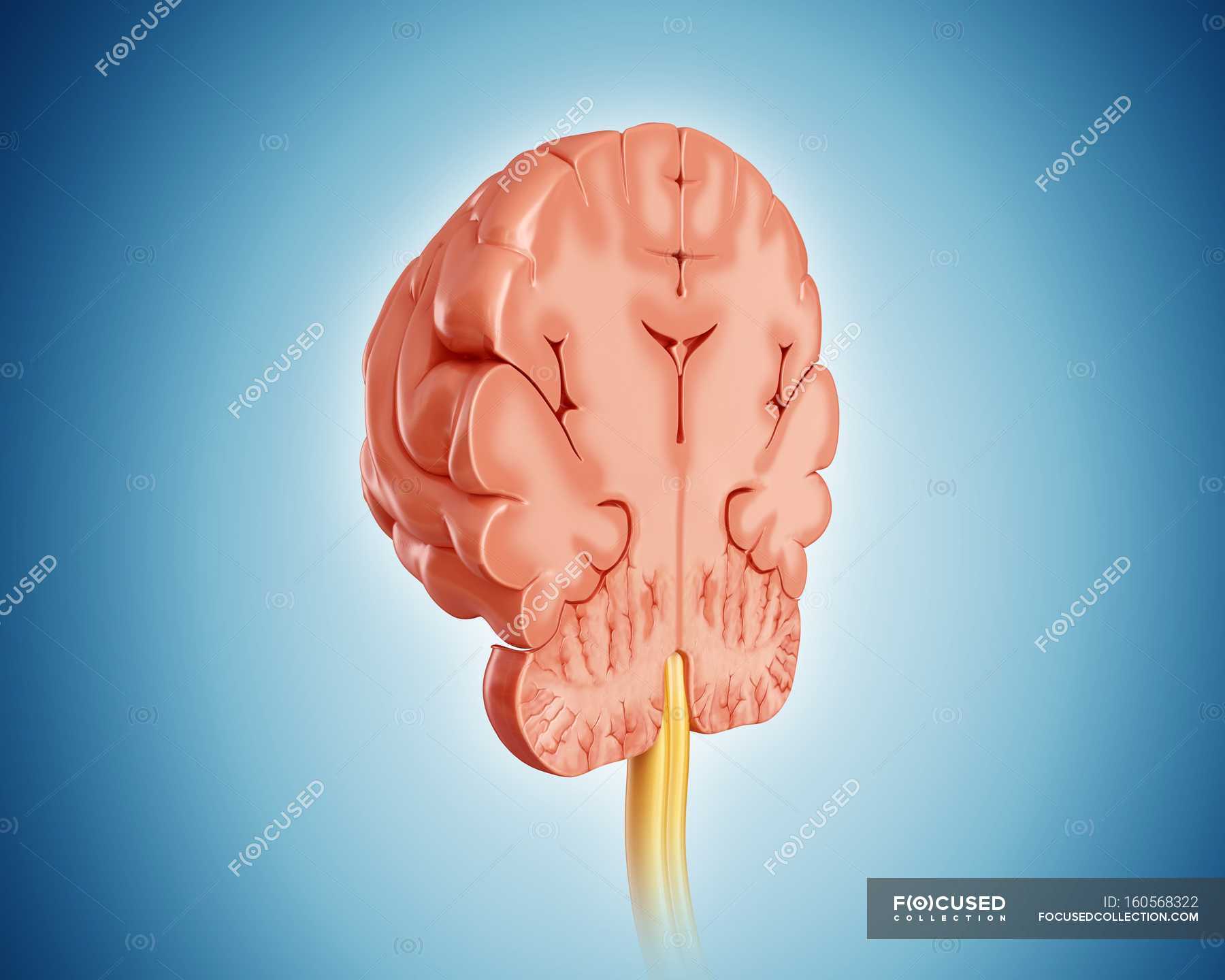
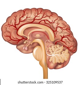
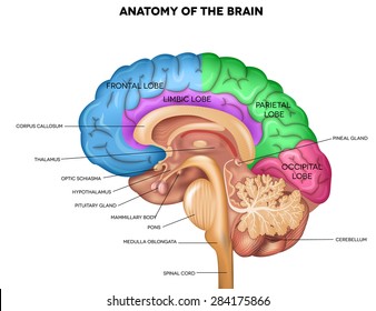
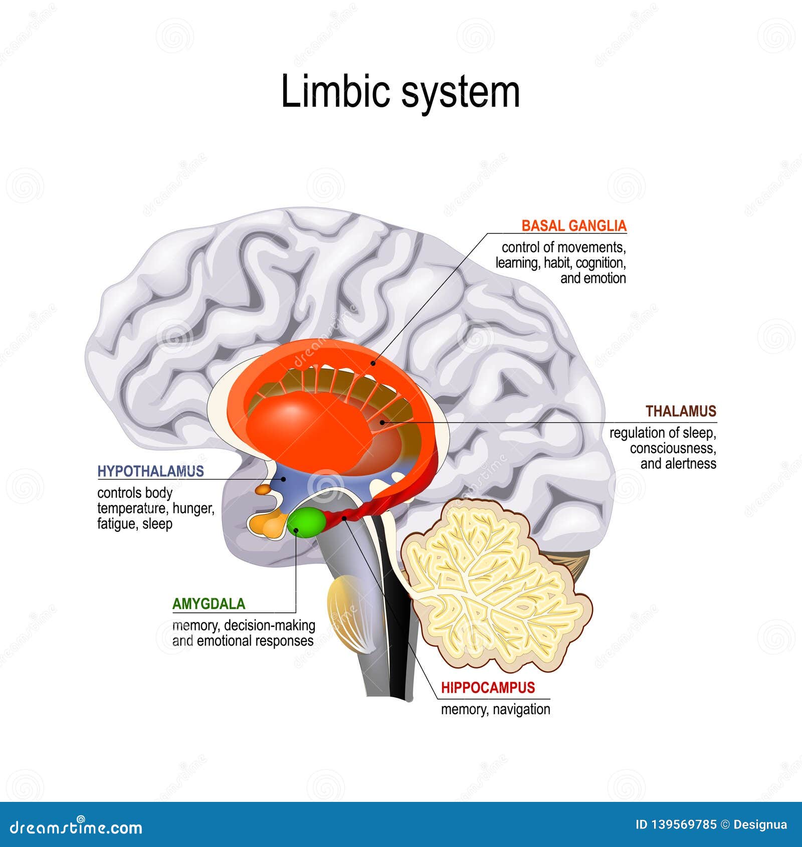
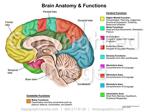
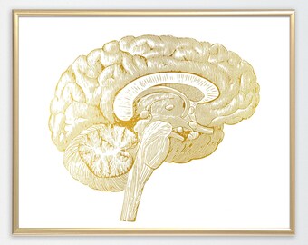
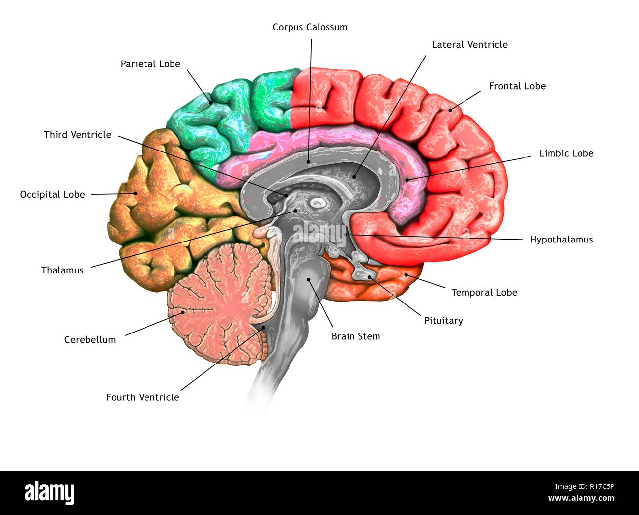
41 brain cross section diagram
en.wikipedia.org › wiki › Pia_materPia mater - Wikipedia The section of the pia mater enveloping the brain is known as the cranial pia mater. It is anchored to the brain by the processes of astrocytes, which are glial cells responsible for many functions, including maintenance of the extracellular space. testbook.com › learn › venn-diagram-reasoningVenn Diagram Reasoning, Key Concepts, Solved Examples, More! Nov 26, 2020 · Types of Venn Diagram As now we know what consists of the questions related to the Venn Diagram reasoning section. Let us see the various types of questions that may come one by one below. 1. Basic Relation. In this type of Venn diagram reasoning, general relations will be given and candidates need to find the best Venn Diagram for those ...
› scitable › knowledgeCoastal Processes and Beaches | Learn Science at Scitable An idealised cross-section of a wave-dominated beach system consisting of the swash zone which contains the subaerial or 'dry' beach (runnel, berm, and beach face) and is dominated by swash ...
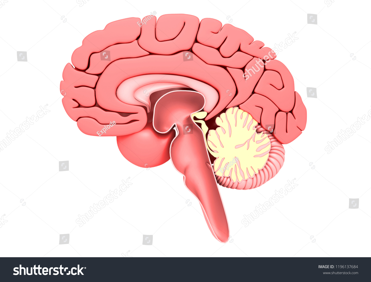
Brain cross section diagram
pubs.rsna.org › doi › fullPatterns of Contrast Enhancement in the Brain and Meninges ... Mar 01, 2007 · Contrast material enhancement for cross-sectional imaging has been used since the mid 1970s for computed tomography and the mid 1980s for magnetic resonance imaging. Knowledge of the patterns and mechanisms of contrast enhancement facilitate radiologic differential diagnosis. Brain and spinal cord enhancement is related to both intravascular and extravascular contrast material. Extraaxial ... healthjade.com › human-earHuman Ear Anatomy - Parts of Ear Structure, Diagram and Ear ... Note: a) Cross section of the cochlea. (b) The spiral organ and the tectorial membrane. (b) The spiral organ and the tectorial membrane. Anterior to the vestibule is the cochlea, a bony spiral canal that resembles a snail’s shell and makes almost three turns around a central bony core called the modiolus. en.wikipedia.org › wiki › BrainstemBrainstem - Wikipedia The brainstem (or brain stem) is the posterior stalk-like part of the brain that connects the cerebrum with the spinal cord. In the human brain the brainstem is composed of the midbrain, the pons, and the medulla oblongata.
Brain cross section diagram. › dishwasher-repair-6Dishwasher Wiring Diagram, Schematic & Cycle Not Advancing Either way, you must remove the door panel to get to it as described in section 5-2. If you already know how to read a wiring diagram, you can skip this section. Each component should be labelled clearly on your diagram. Look at figure 6-A. The symbols used to represent each component are pretty universal. en.wikipedia.org › wiki › BrainstemBrainstem - Wikipedia The brainstem (or brain stem) is the posterior stalk-like part of the brain that connects the cerebrum with the spinal cord. In the human brain the brainstem is composed of the midbrain, the pons, and the medulla oblongata. healthjade.com › human-earHuman Ear Anatomy - Parts of Ear Structure, Diagram and Ear ... Note: a) Cross section of the cochlea. (b) The spiral organ and the tectorial membrane. (b) The spiral organ and the tectorial membrane. Anterior to the vestibule is the cochlea, a bony spiral canal that resembles a snail’s shell and makes almost three turns around a central bony core called the modiolus. pubs.rsna.org › doi › fullPatterns of Contrast Enhancement in the Brain and Meninges ... Mar 01, 2007 · Contrast material enhancement for cross-sectional imaging has been used since the mid 1970s for computed tomography and the mid 1980s for magnetic resonance imaging. Knowledge of the patterns and mechanisms of contrast enhancement facilitate radiologic differential diagnosis. Brain and spinal cord enhancement is related to both intravascular and extravascular contrast material. Extraaxial ...















Comments
Post a Comment