41 onion epidermal cell labeled diagram
Because of their simple structure and.onion epidermal cell labeled onion cell diagram Wide collections of all kinds of labels pictures online. Make your work easier by using a label. Happy Labeling! Labels are a means of identifying a product or container through a piece of fabric, paper, metal or plastic film onto which information about them is printed. The information can be. Annotated ... The onion epidermis cell is the only cell that has a cell wall. In addition, it is the only cell that has a chloroplast, where photosynthesis can happen. The cheek epithelium cell is the only one that has centrioles, the barrel-shaped organelle that is responsible for helping organize chromosomes during cell division.
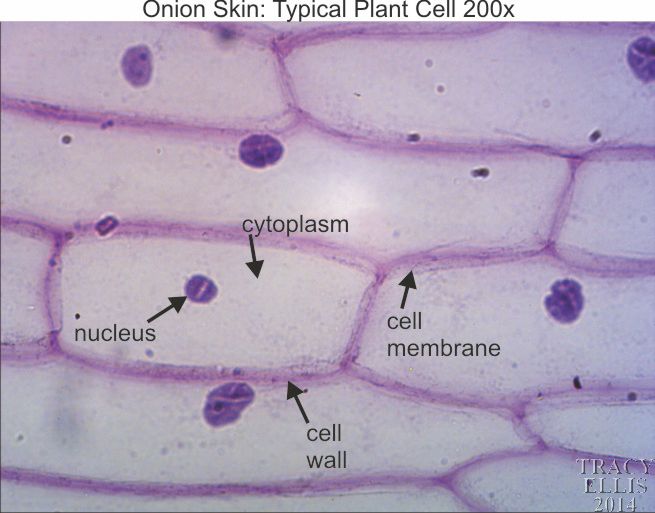
Onion Cell. An onion is a multicellular (consisting of many cells) plant organism.As in all plant cells, the cell of an onion peel consists of a cell wall, cell membrane, cytoplasm, nucleus and a large vacuole. The nucleus is present at the periphery of the cytoplasm. The vacuole is prominent and present at the centre of the cell. It is surrounded by cytoplasm. The presence of a cell wall and ...

Onion epidermal cell labeled diagram
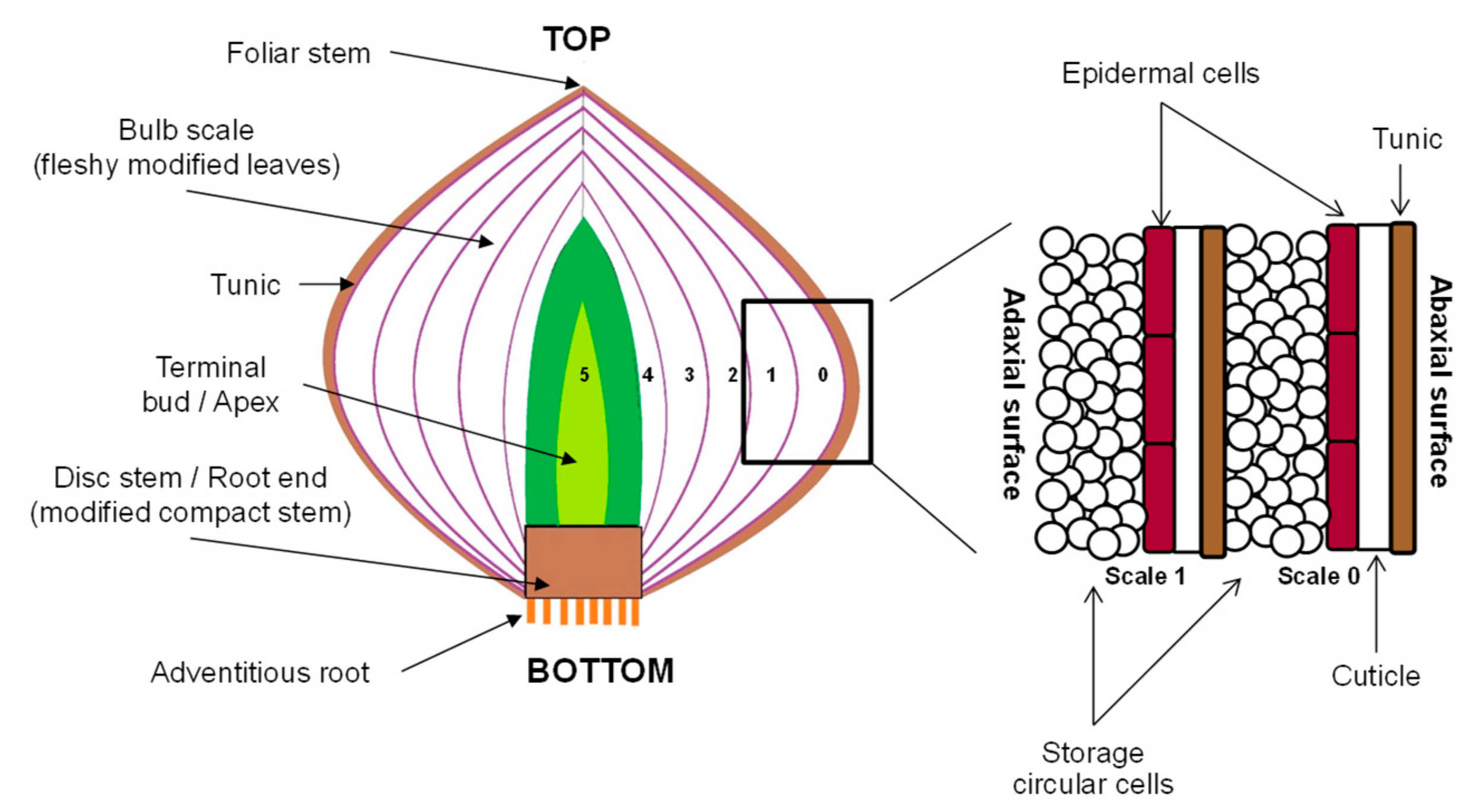
Onion Epidermal Cell Labeled. Wide collections of all kinds of labels pictures online. Make your work easier by using a label. Happy Labeling! Labels are a means of identifying a product or container through a piece of fabric, paper, metal or plastic film onto which information about them is printed. The information can be in the form of hand ... Printable Animal Cell Diagram - Labeled, Unlabeled, and Blank; 您是否在找: labeled和labelled labelme vim下一页 labeled mute dispute和argue区别 be ... Onion Epidermis. Onion at 100× Onion at 400× Peeling Onion Epidermis Each layer of an onion consists of a thick, fleshy layer sandwiched between two shiny, transparent, membranous, epidermal layers. Peel a small piece of the transparent epidermis from a layer of an onion (NOTE: YOU DO NOT WANT the whole, thick, fleshy part, just the ...
Onion epidermal cell labeled diagram. The epidermal cells of onions provide a protective layer against viruses and fungi that may harm the sensitive tissues. Because of their simple structure and transparency they are often used to introduce students to plant anatomy or to demonstrate plasmolysis. The clear epidermal cells exist in a single layer and do not contain chloroplasts, because the onion fruiting body (bulb) is used for ... Onion epidermal cell labeled diagram. Cell diagram 1 onion benjamin himme. The microscope is the device which helps scientists observe how the onion epidermal layer looks like. Label the cell wall and chloroplasts. Onion cells are among the most common choices for cell studies in early biology classes. Easily obtained inexpensive they offer samples with no difficult technique required. Labeled ... Scytek Alarm Wiring Diagram. Vehicle security system with remote start with seperate lock/unlock buttons (31 pages). Car Alarm Scytek electronic Astra 7T7 Series Product Manual. If the system is triggered by the doors, or hood/trunk, the system will alarm for 45 seconds. . Diagram below shows the onion cell when viewed under a microscope. 3. Fig 1.1.3.jpg (353 KB) Fig 1.1.3: Onion epidermal cells stained with iodine seen under ...
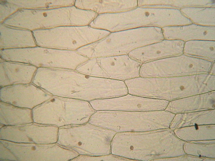
Find onion cell stock images in HD and millions of other royalty-free stock photos, ... Onion epidermis with large cells under light microscope. An epidermal onion cell diagram includes components such as citoplasm, a round nucleus and a cell wall. The microscope is the device which helps scientists observe how the onion epidermal layer looks like. Although at the surface an onion has dried protective leaf, inside of it, things change. The onion cell diagram shows that in the middle of. Having observed the onion cell under the microscope, students will be able to learn the differences between animal and plant cells in addition to the function of the different parts of the cell. Learn about Onion Root Tip Mitosis. View Epidermal Cells. Check out other microscope experiment viewing Cheek Cells, Cork Cells or Sugar Crystals as well and have fun looking at Leaf Structure under ... Epidermal Cells in Onion. Epidermal cells of onions are very simple. As a result, the epidermal tissue has become the ideal model through which students are introduced to the morphology/anatomy of plant cells. Epidermal cells of onions also have well-defined shapes that may appear rectangular or square (or as elongated hexagonal) under the microscope. When viewed under the microscope, it is ...
The figure given below, shows the epidermal cells of an onion bulb. This cell was then transferred to a drop of sugar solution. (i) Draw a well labelled diagram of the epidermal cell as it would appear after immersion in a strong sugar solution. (ii) What scientific term is used for the changes as shown in (i) above ? (iii) What should be done to restore the cell back to its original condition ... GTAC Osmosis in red onion Page 2 of 2 Method - onion epidermal peel a) Collect a segment of red onion and all other materials. Label 2 microscope slides: 'dH2O' and 'salt solution'. b) Peel a thin layer of epidermis from a red surface of the onion. c) Place it on the slide. The figure given below shows the epidermal cells of an onion bulb. This cell was then transferred to a drop of sugar solution. (i) Draw a well labelled diagram of the epidermal cells as it would appear after immersion in a strong sugar solution. (ii) What scientific term is used for the changes as shown in (i) above? All living organisms are made up of cells. The shape, size and the number of these units vary in organisms. The three major components of a cell are the cell membrane, cytoplasm and nucleus. In a plant cell, a cell wall surrounds the cell membrane. Procedure: 1. Take an onion and remove its outermost peel. 2.
Plant Cells And Tissues
Onion cell 40x labeled diagram bsa positive ground wiring diagram usb to ethernet wiring diagram att phone box wiring diagram blank mouth diagram ignition wiring. The nucleus of an onion epidermal cell. Onion epidermis at 40x iodine stain. Onion cell 40x labeled diagram the cellulose present in the cell walls forms clearly defined tiles. Onion ...

Observing Onion Peel Epidermal Cells Under Microscope Best Demo Biology Youtube
Onion Cell. An onion is a multicellular (consisting of many cells) plant organism. As in all plant cells, the cell of an onion peel consists of a cell wall, cell membrane, cytoplasm, nucleus and a large vacuole. The nucleus is present at the periphery of the cytoplasm. The vacuole is prominent and present at the centre of the cell.
Onion Cells High Resolution Stock Photography And Images Alamy
7. Make a wet mount of the onion tissue you just rinsed using 2 or 3 drops of DISTILLED WATER on the onion tissue then install cover slip. Watch the cells for approximately 2-3 minutes or longer as you again survey the ENTIRE onion tissue on LOW POWER. You should see changes within many of the cells initially near the perimeter of the onion tissue.

Learning By Questions
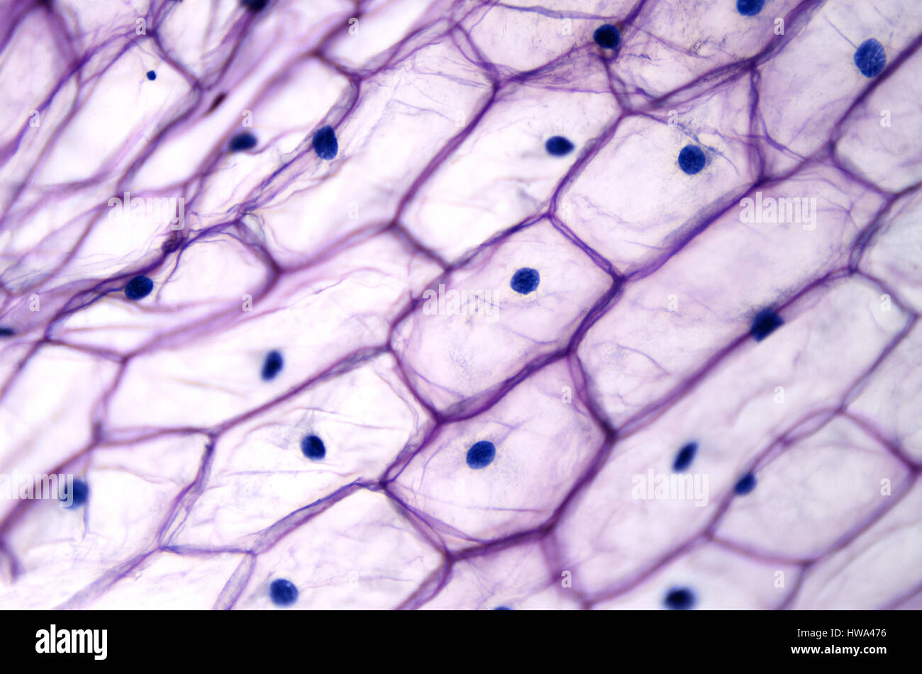
Onion epidermis, at 100X, iodine stain. Onion epidermal cells, iodine stain, 400X. The nucleus of an onion epidermal cell, 1000X magnification ...

Edwin Joseph Edwinj0123 Profile Pinterest
Diagram below shows the onion cell when viewed under a microscope. 3. Fig 1.1.3.jpg (353 KB) Fig 1.1.3: Onion epidermal cells stained with iodine seen under ...
Cell And Microscopes Email Quiz Homework Onion Cell Diagrams
Let's try how to make onion cell...
2
The epidermis in typical hydrophyte has an extremely thin cuticle, and the thin cellulose walls permit ready absorption from the surrounding water. Generally the chloroplasts are found in epidermal cells of leaves, especially when the leaves are very thin; these chloroplasts utilize the weak light under water for photosynthesis.

Onion Epidermis Under Light Microscope Purple Colored Large Microscopic Photography Epidermis Skin Cells
Video explaining how to draw a biological diagram showing cell detail.
1
Onion Epidermis With Large Cells Under Microscope Stock Image. Lab Drawings Biology For Life. Measuring Cells Ppt Video Online Download. Onion Cell Mitosis Stages. Onion Cell Diagram Labelled Wiring Schematic Diagram. Draw Diagram Of Onion Peel As It Is Seen Through Microscope From.
Mic Uk
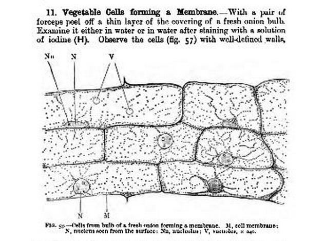
The skin (or epidermis) between the dormant leaves of an onion are a single cell thick, and serve as a classic representation of the internal structure of plant cells. In fact, the term "cell" came from a pioneer of microscopic biology, Robert Hooke, while looking at epidermal onion cells under a microscope.
School Science How To Prepare An Onion Cell Slide Wikibooks Open Books For An Open World
onion epidermal cell labeled onion cell diagram 02. Wide collections of all kinds of labels pictures online. Make your work easier by using a label. Happy Labeling! Labels are a means of identifying a product or container through a piece of fabric, paper, metal or plastic film onto which information about them is printed.
The Figure Given Below Shows The Epidermal Cells Of An Onion Bulb This Cell Was Then Transferred To A Drop Of Sugar Solution Sarthaks Econnect Largest Online Education Community
(*Hint — focus on cells located near the edge of the coverslip where you added the stain. These cells will get stained first.) 3. Observe the onion cells under low power. 4. Draw two adjacent onion cells. Identify and label the cell wall, cell membrane, nucleus, and nucleolus, if visible. 5. Removethe slide from the stage and save itfor later.

Sketch The Onion Peel Cell As Seen Under The Microscope Label The Parts Such As The Cell Wall Brainly In
Onion Cell Lab Report By : Nawaf Almalki Introduction: Many things that are viewed using a microscope, particularly cells, can appear quite transparent under the microscope. The internal parts of the cells, the organelles, are so transparent that they are often difficult to see. Biologists have developed a number of stains that help them see the cells and their organelles by adding color to ...
Foods Free Full Text Structural Changes Induced By Pulsed Electric Fields Increase The Concentration Of Volatiles Released In Red Onion Allium Cepa L Var Red Pearl Bulbs Html
Dra A Full Labeled Diagram Of An Epidermal Cell With Chegg Com. Solved Ve Ds E Ele 4 The Diagram Alongside Represents A Layer Of Epidermal Cells Showing A Fully Grown Root Hair Study The Diagram And Answer The Questions That Follow A Name. 1 The Diagram Below Represents A Layer Of Epidermalcells Showing A Fully Grown Root Hair Study Brainly In.

Cell Microscope Investigation Ppt Video Online Download
Epidermal Onion Cells Under A Microscope Plant Cells Appear Polygonal From The Cell Diagram Plant Cell Diagram Plant Cell . T S Older Root Biologi . The Cell An Image Library Image Cil 39065 Mitosis Cell Processes Mitosis Activity . 62100604d 6wr Fundamental Photographs The Art Of Science Mitosis Mitosis Activity Biology Units . This Image Or Other Media File Is In The Public Domain Because ...

Ruzivo Digital Learning
To demonstrate the phenomenon of plasmolysis. Tradescantia leaf, water, sugar solution, slides, cover glass, microscope, blade. 1. From the lower surface of the leaf of Tradescantia, peel off small segments of epidermis by a blade. 2. Put few peelings on a slide, mount in a drop of water, put a cover glass and study under microscope. 3. Mount ...

Onion Epidermis
Light Micrograph (LM) of onion skin cells, magnification x 600 - Stock Image ... Clear epidermal cells of an onion, Allium cepa, in a single layer.

A Thin Strip Of Epidermal Cells From The Fleshy Scales Of An Onion Bulb Was Examined In A Drop Of Water Under A Microscope Biology Shaalaa Com
Onion Epidermis. Onion at 100× Onion at 400× Peeling Onion Epidermis Each layer of an onion consists of a thick, fleshy layer sandwiched between two shiny, transparent, membranous, epidermal layers. Peel a small piece of the transparent epidermis from a layer of an onion (NOTE: YOU DO NOT WANT the whole, thick, fleshy part, just the ...

Lesson 3 Onion Dissection Look At The Plant Cells Rs Science
Printable Animal Cell Diagram - Labeled, Unlabeled, and Blank; 您是否在找: labeled和labelled labelme vim下一页 labeled mute dispute和argue区别 be ...

Observing Onion Cells Under The Microscope Microscope Club
Onion Epidermal Cell Labeled. Wide collections of all kinds of labels pictures online. Make your work easier by using a label. Happy Labeling! Labels are a means of identifying a product or container through a piece of fabric, paper, metal or plastic film onto which information about them is printed. The information can be in the form of hand ...

Plant Cell Parts Flashcards Quizlet
Solved Fig 2 7 Stained Cells Of Fleshy Onion Allium Cepa Leaves Observed Under Microscope 10 Um Course Hero
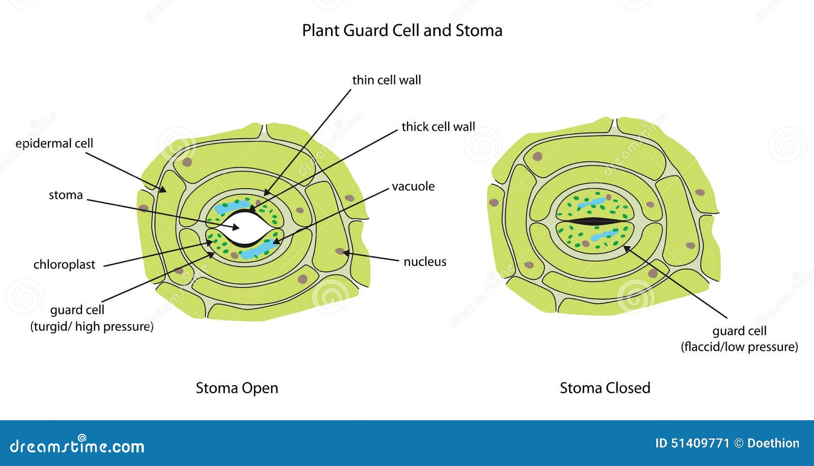
Plant Guard Cells With Stoma Fully Labeled Stock Illustration Illustration Of Epidermal Stomata 51409771
If Onion Peel Cells And Cheek Are Observed Through A Microscope What Are Two Major Differences That The Observer Is Likely To Find Quora

Onion Epidermis

Under The Micrsocope Onion Cell 100x 400x Youtube
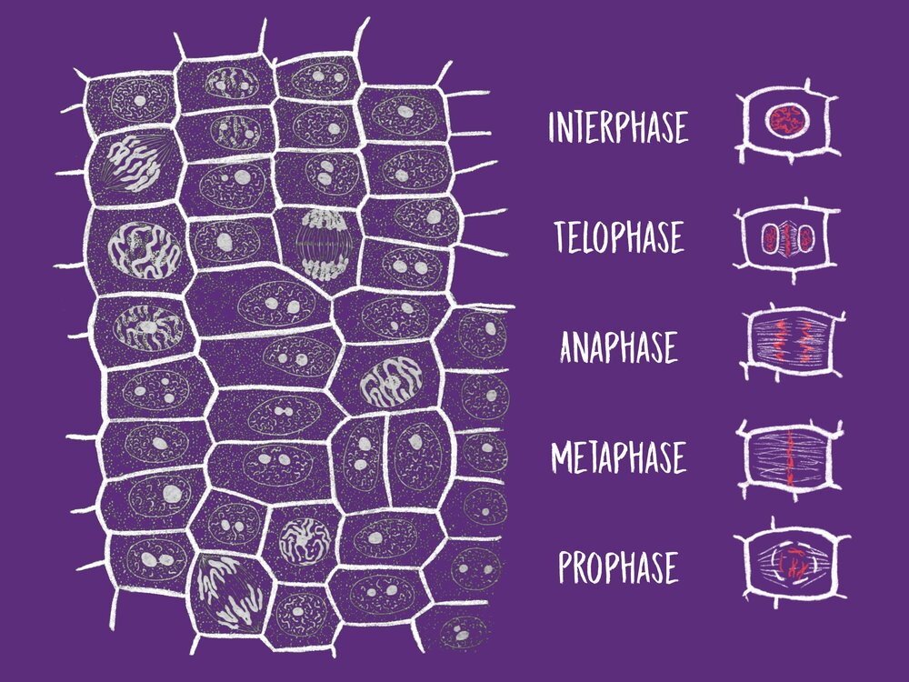
Mitosis In Onion Root Tips Dataclassroom

Onion Cell Labelling Diagram Quizlet
Mic Uk How Many Onion Skins Are There

Lesson 3 Onion Dissection Look At The Plant Cells Rs Science
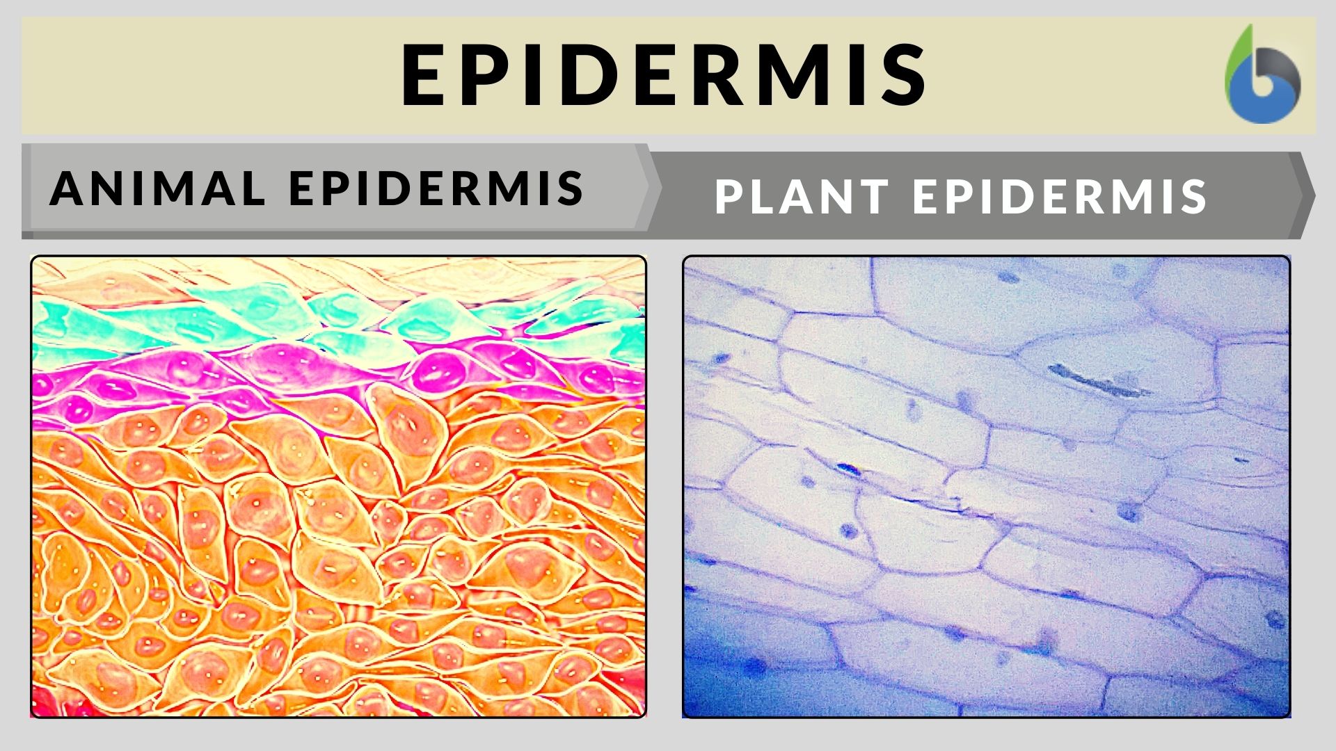
Epidermis Definition And Examples Biology Online Dictionary

Living And Learning August 2010
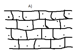
The Following Diagram Shows Cells Of Onion Peel Label Class 11 Biology Cbse

Schematic Of The Onion Epidermal Cells A Obtaining The Onion Download Scientific Diagram

Labeling Of Microfilaments In Onion Bulb Scale Epidermis Cells Download Scientific Diagram

Observing Onion Cells Under The Microscope Microscope Club

Tomato Cells Under Microscope

Mitochondrial Fusion In Onion Bulb Epidermal Cells Timecourse Download Scientific Diagram

Module 1 Secondary Science Biology View As Single Page
Onion Skin 200x Dissection Connection

Post Lab Questions Microscopy


Comments
Post a Comment