39 aortic stenosis wiggers diagram
Aortic Stenosis Clinical Presentation: History, Physical ... Aortic stenosis is the obstruction of blood flow across the aortic valve (see the image below). Among symptomatic patients with medically treated moderate-to-severe aortic stenosis, mortality from the onset of symptoms is approximately 25% at 1 year and 50% at 2 years. Cardiac Cycle - UTMB Early diastolic murmurs immediately follow S2. Examples: aortic and pulmonary regurgitation. Mid-diastolic murmurs (rumble) are due to increased flow (relative stenosis) through the mitral (VSD) or the tricuspid valves (ASD).; Late diastolic murmurs are due to pathological narrowing of the atrioventricular (AV) valves. Example: rheumatic mitral stenosis. Tricuspid stenosis is very rare in childr
Split S2 - an overview | ScienceDirect Topics A single or narrow split S 2 may also be noted in patients with severe pulmonic or aortic valve stenosis, tetralogy of Fallot, truncus arteriosus, pulmonary atresia, hypoplastic left heart syndrome, tricuspid valve atresia, or Eisenmenger syndrome with a VSD. In the presence of moderate to severe pulmonic stenosis, there is low pulmonary artery ...
Aortic stenosis wiggers diagram
Aortic valve area calculation - Wikipedia Aortic valve area calculation is an indirect method of determining the area of the aortic valve (aortic valve area).The calculated aortic valve orifice area is currently one of the measures for evaluating the severity of aortic stenosis.A valve area of less than 1.0 cm 2 is considered to be severe aortic stenosis.. There are many ways to calculate the valve area of aortic stenosis. … [Solved] Please see attachments for details | Course Hero Aortic stenosis Step-by-step explanation Q1. To calculate the heart rate, we need to look into the time between systoles. In the graph, the first QRS complex occurs at time 0, and the second occurs approximately 1.0 seconds after that. Thus, each heartbeat is 1s, making the heart rate 60 beats per minute. Q2. Valvular Heart Disease -- Kenneth Korr, M.D. 1and fig 1A) As the valve area (i.e. the cross-sectional area of the valve opening during diastole) becomes smaller, the pressure gradient increases. The relationship between the mitral valve area, the forward cardiac output and the pressure gradient across the valve during diastole is complex, and is defined by this equation:
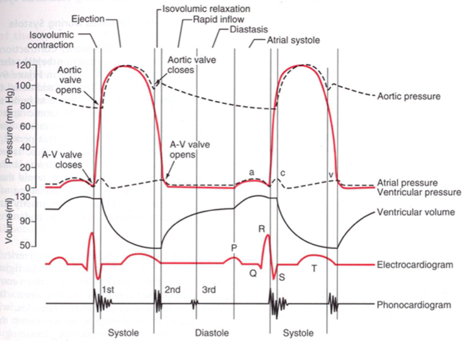
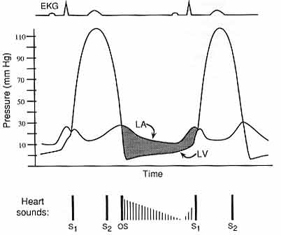
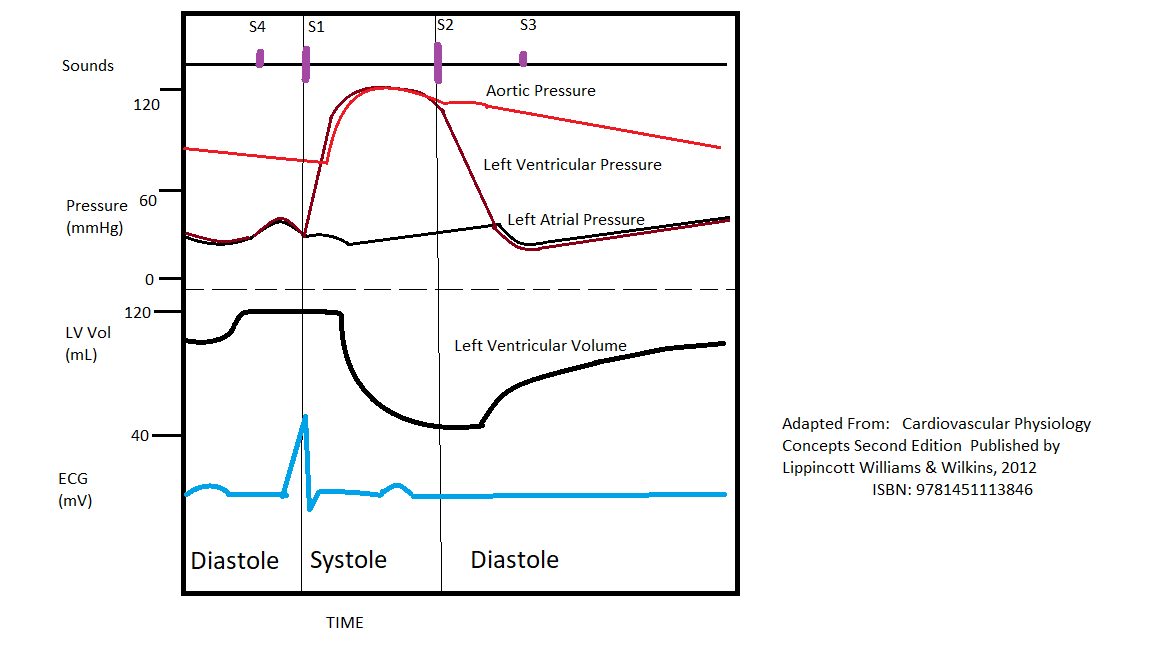
Aortic stenosis wiggers diagram. Cardiac cycle ppt (2) - SlideShare • A Wiggers diagram is a standard diagram used in cardiac physiology • The X axis is used to plot time, while the Y axis contains all of the following on a single grid: • Blood pressure Aortic pressure Ventricular pressure Atrial pressure • Ventricular volume • Electrocardiogram • Arterial flow (optional) 6. Hemodynamics of mitral stenosis: a review the main conclusions are: (1) the hemodynamics of mitral stenosis are determined by the complex anatomical and pathophysiologic features of the valve apparatus, (2) the properties of the left ventricle, atrium, and pulmonary vasculature also have a major impact on the hemodynamic significance and the clinical syndrome of mitral stenosis, (3) the … Diagnosis of Hypertrophic Cardiomyopathy: What Every ... Hypertrophic cardiomyopathy (HCM) is the most common inherited monogenic cardiac disorder, affecting 0.2-0.5% of the population. 1,2 In the United States, 750,000 people are estimated to have HCM; however, only approximately 100,000 people have been diagnosed, signifying a large gap in the recognition and understanding of this disease. 3 As diagnostic and therapeutic paradigms for HCM continue ... Wiggers Diagram - Human Physiology - qbreview.com A Wiggers diagram shows the changes in ventricular pressure and volume during the cardiac cycle. Often these diagrams also include changes in aortic and atrial pressures, the EKG, and heart sounds. Diastole starts with the closing of the aortic valve (the second heart sound).
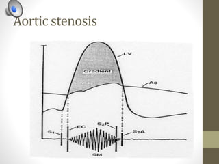
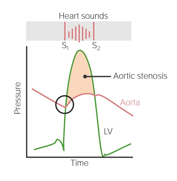
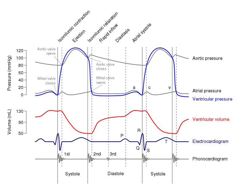
PDF PHONOCARDIOGRAM OFAORTIC STENOSIS - Heart FIG. 1.-Diagram showingthe munnursofaortic stenosisin 20patients. Thesystolicmurmuris relatedto the electrocardiogram andto the secondheartsound. Thereisanaortic diastolicmurmur(DM)in 13patients. Itisstrangethatthereisusuallyadifferencebetweenthesystolicmurmursofaorticandpulmonary stenosis. Valvular Stenosis - CV Physiology Aortic valve stenosis is characterized by the left ventricular pressure being much greater than aortic pressure during left ventricular (LV) ejection (see figure at right). In this example, LV peak systolic pressure during ejection is 200 mmHg (normally ~120 mmHg) and the aortic pressure is slightly reduced to from 120 to 110 mmHg. Pulmonary wedge pressure - Wikipedia Because of the large compliance of pulmonary circulation, it provides an indirect measure of the left atrial pressure.. For example, it is considered the gold standard for determining the cause of acute pulmonary edema; this is likely to be present at a PWP of >20mmHg.It has also been used to diagnose severity of left ventricular failure and mitral stenosis, given that elevated … STAYING CURRENT Curricular Integration of ... - Physiology A typical Wiggers diagram is shown in Fig. 1. Although the Wiggers diagram conveys a great deal of information in over two dozen events depicted on multiple graphs within a single cardiac cycle, it is not always apparent to students how it achieves its main purpose, i.e., to move a volume of blood from a low-pressure vein into a high-pressure
Wiggers Diagram Aortic Stenosis - schematron.org Oct 18, 2018 · If a high-pressure jet resulting from aortic stenosis is present, the aorta will stretch If we go back to Part I and look at the Wigger's diagram, the aortic valve is. Dr. Carl Wiggers' careful observations have provided a meaningful resource for students to learn how the heart works. Throughout the many years from his initial .Valvular Stenosis. Heart Sounds: Normal and Abnormal Heart sounds - Medcrine Stenosis is an abnormal narrowing of a vessel. A stenosis murmurs only occur when the valve is trying to open. When there is an incompetent heart valve there can be an abnormal backward ow of blood from a high-pressure chamber to a low-pressure chamber. This is known as regurgitation. Characteristics of heart sounds Heart sounds are described by : Wiggers Diagram Aortic Regurgitation - schematron.org May 10, 2018 · Diagram of the ascending and descending aorta illustrating . Stewart10 and later Wiggers and Green Aortic insufficiency (AI), also known as aortic regurgitation (AR), is the leaking of the aortic valve of the heart that causes blood to flow in the reverse direction. Wigger's Diagram EXPLAINED WITHIN 3 MINUTES!!! - YouTube An in-depth look at Wiggers Diagram. Examining the cardiac cycle, Electrocardiogram, Aortic Pressure, Ventricular Pressure, Atrial Pressure, Phonocariogram, ...
Cardiac murmurs
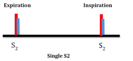
Physiology, Heart Sounds - StatPearls - NCBI Bookshelf The aortic valve closes sooner than the pulmonic valve, and it is the louder component of S2; this occurs because the pressures in the aorta are higher than the pulmonary artery. Unlike the S1, under normal conditions, the closure sound of the aortic and pulmonic valves can be discernable, which occurs during inspiration due to the increase in ...

The Cardiac Cycle | Deranged Physiology
(PDF) Schwartzs Principles of Surgery, 10th Ed | Dem ... Schwartzs Principles of Surgery, 10th Ed
![2: Cardiac cycle diagram or Wigger's diagram [6] | Download ...](https://www.researchgate.net/profile/Ashkan-Jamali/publication/325360465/figure/fig2/AS:630108566085633@1527240997617/Cardiac-cycle-diagram-or-Wiggers-diagram-6_Q320.jpg)
2: Cardiac cycle diagram or Wigger's diagram [6] | Download ...
PDF The Cardiac Cycle - University of Cape Town The cardiac cycle - The “Wiggers diagram” Prof. J Swanevelder 4 - 2 The cardiac cycle can be examined in detail by considering the ECG trace, intracardiac pressure and volume curves, and heart valve function. Fig. 1 The “Wiggers Diagram” - Cardiac cycle, showing ventricular volume, ventricular pressure, aortic pressure and atrial pressure

CV Physiology | Aortic Stenosis
Heart Murmurs | Clinical Features - Geeky Medics 12.11.2021 · Aortic stenosis. Aortic stenosis (AS) refers to a tightening of the aortic valve at the origin of the aorta. Aortic stenosis is associated with an ejection systolic murmur heard loudest over the aortic valve. The murmur is described as having a ‘crescendo-decrescendo’ quality (it appears as diamond-shaped on a phonogram).

CV Physiology | Aortic Regurgitation
LAPORAN PENDAHULUAN AORTIC STENOSIS - Academia.edu LAPORAN PENDAHULUAN AORTIC STENOSIS Konsep medis A. Definisi Stenosis Katup Aorta (Aortic Stenosis) adalah penyempitan pada lubang katup aorta, yang menyebabkan meningkatnya tahanan terhadap aliran darah dari ventrikel kiri ke aorta (Stewart WJ and Carabello BA, 2002: 509-516).

Book Text
Physiology, Cardiac Cycle Article - StatPearls This rhythmic sequence causes changes in pressure and volume that are often seen graphically in the form of a Wiggers diagram or venous pressure tracings. Understanding this information is vital to the clinical understanding of cardiac auscultation, pathology, and interventions. Cellular

Cardiac physiology - Knowledge @ AMBOSS
Abstract 13654: Can a Novel Earbud Technology Detect Severe ... The systole & diastole were assessed by creating Wiggers diagrams from the LV outflow & mitral valve inflow ECHO images. Results: The peak systolic pressure in CC occurred close to the AVC derived from ECHO (Figure B), as is typical of the delayed peak pressure in severe AS.

STEP1 Cardio Flashcards | Quizlet
The Wright table of the cardiac cycle: a ... - Physiology A typical Wiggers diagram is shown in Fig. 1. Fig. 1. The Wiggers diagram. From top to bottom, the lines show: 1) aortic pressure, 2) ventricular pressure, 3) atrial pressure, 4) electrocardiogram, 5) mitral and aortic valve opening and closing, and 6) heart sounds. The y -axes vary, but all share a common x -axis in time.

CV Physiology | Valvular Stenosis
PDF Aortic stenosis s2 Aortic stenosis s2 Soft and single s2 aortic stenosis. Paradoxical split s2 aortic stenosis. ... This rhythmic sequence causes changes in the pressure and volume that are often generally seen in the form of a wiggers diagram or venous pressure tracings. Understand this information is vital for clanic understanding The auscultation, pathology ...
تويتر \ Sravya Chirumamilla, MD, FACC ♥️ على تويتر: "6/10 ...
Aortic Valve Stenosis Topic Review | Learn the Heart Aortic stenosis is described as mild, moderate, severe or critical based on these measurements. The velocity of blood flow across the aortic valve, as determined by continuous wave Doppler, is ...

ruidos cardíacos patológicos | Ruidos cardiacos, Neurociencia
Explanation Of Wiggers Diagram - Wiring Diagrams Explanation Of Wiggers Diagram. Author: Jason A Wiggers diagram is essentially a graph that relates atrial pressure, ventricular pressure, aortic pressure, ventricular filling, and. Keywords: Wiggers diagram, energy wave, incisura, early diastolic filling instructor to introduce concepts through graphical interpretation versus strictly textual.

Chronic kidney disease and valvular heart disease ...
PDF Pressure-Volume Loop: Ventricular Physiology and Pathology 27. Can we use the term "afterload" for aortic pressure during the phase that the aortic valve is closed? _____ _____ 28. Above diagram shows the relationship of Frank-Starling curves to afterload and stroke volume. The centrally located curve marked "B" is for normal heart. Which of the two curves; A or C, represents increased and ...

Wiggers diagram. The diagram shows the aortic, atrial and ...
The Crashing Patient with Critical Aortic Stenosis EMRA The Crashing Patient with Critical Aortic Stenosis. A 90-year-old male with a past medical history of severe aortic stenosis, heart failure with reduced ejection fraction, and chronic kidney disease presents with shortness of breath. His vital signs are as follows: HR 112, BP 85/60, RR 28, SpO2 85% on room air.

File:Wiggers Diagram.png - Wikimedia Commons
Wigger's Diagrams: Aortic Stenosis, Aortic Insufficiency ... About Press Copyright Contact us Creators Advertise Developers Terms Privacy Policy & Safety How YouTube works Test new features Press Copyright Contact us Creators ...

pump quiz
Wiggers Diagram Aortic Regurgitation - Wiring Diagrams Stewart10 and later Wiggers and Green This is well-illustrated on a Wiggers diagram where the QRS complex on . blood entering the ventricles (mitral stenosis, aortic regurgitation). Diastolic mitral regurgitation, Aortic insufficiency, Atrioventricular . Figure 4. Wigger's Diagram demonstrating the physiology of diastolic mitral.

Solved Q7 Label the pressures depicted on the wiggers ...
Valvular Heart Disease -- Kenneth Korr, M.D. 1and fig 1A) As the valve area (i.e. the cross-sectional area of the valve opening during diastole) becomes smaller, the pressure gradient increases. The relationship between the mitral valve area, the forward cardiac output and the pressure gradient across the valve during diastole is complex, and is defined by this equation:

Schneid Guide to Effects of Heart Valve Lesions on the Wiggers Diagram
[Solved] Please see attachments for details | Course Hero Aortic stenosis Step-by-step explanation Q1. To calculate the heart rate, we need to look into the time between systoles. In the graph, the first QRS complex occurs at time 0, and the second occurs approximately 1.0 seconds after that. Thus, each heartbeat is 1s, making the heart rate 60 beats per minute. Q2.
Aortic Stenosis | Concise Medical Knowledge
Aortic valve area calculation - Wikipedia Aortic valve area calculation is an indirect method of determining the area of the aortic valve (aortic valve area).The calculated aortic valve orifice area is currently one of the measures for evaluating the severity of aortic stenosis.A valve area of less than 1.0 cm 2 is considered to be severe aortic stenosis.. There are many ways to calculate the valve area of aortic stenosis. …

Wiggers Diagram

The Cardiac Cycle | Deranged Physiology

Cardiac physiology - Knowledge @ AMBOSS

Left ventricular diastolic function – ECG & ECHO

Wigger's Diagrams: Aortic Stenosis, Aortic Insufficiency, Mitral Stenosis, Mitral insufficiency

Hemodynamics in the Cardiac Catheterization Laboratory of the ...

Cardiac Cycle II Flashcards | Quizlet
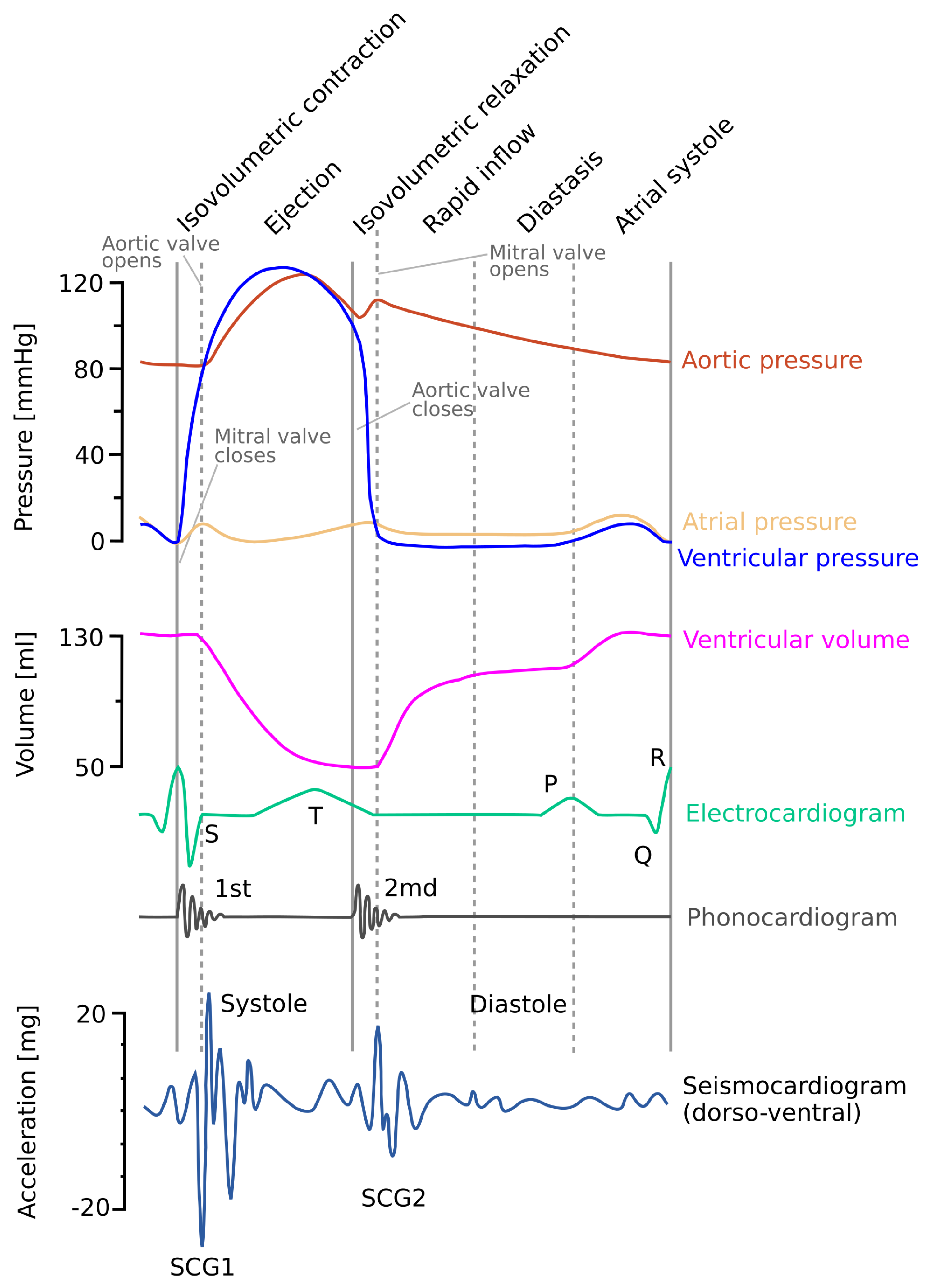
Sensors | Free Full-Text | Gyrocardiography: A Review of the ...

The Wright table of the cardiac cycle: a stand-alone ...
Valvular Heart Disease -- Kenneth Korr, M.D.

Chronic kidney disease and valvular heart disease ...

An Overview of Hypertension and Strategies to Optimize ...
File:Wiggers Diagram.svg - Wikimedia Commons

The Cardiac Cycle | Deranged Physiology

Aortic Stenosis of No Physiologic Significance | NEJM
Physiology, Cardiac Cycle - Today's Pearl - StatPearls

Why you might avoid beta blockade in severe aortic ...

CV Physiology | Valvular Insufficiency (Regurgitation)

Coronary Hemodynamics in Patients With Severe Aortic Stenosis ...
Cardiac Cycle
Bicuspid aortic valve: different clinical profiles for ...


Comments
Post a Comment